Watch Out! Molecular biology requires precision at the nanoscale:
Misconception 1:All DNA in a cell codes for proteins.Correction: In humans, less than 2% of DNA actually codes for proteins! The vast majority is non-coding DNA, which plays crucial roles in regulation and chromosome structure.
Misconception 2:Mutations are always harmful and cause diseases.Correction: Mutations are the raw material for evolution. Many are completely neutral (silent mutations), and some are even beneficial depending on the environment.
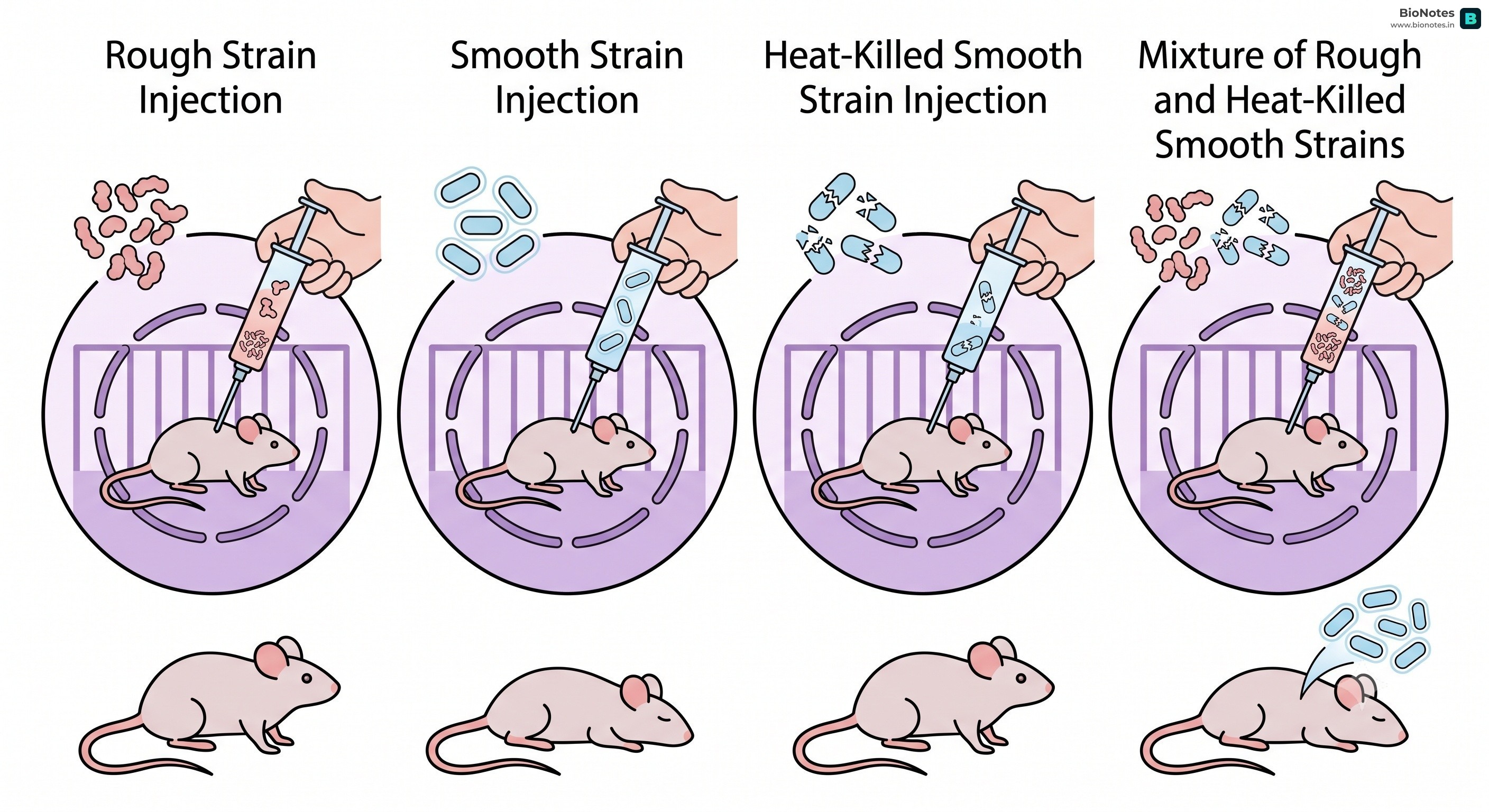
Frederick Griffith worked with Streptococcus pneumoniae (pneumococcus) bacteria and mice.
R strain (rough): Non-virulent, no capsule.
S strain (smooth): Virulent, with capsule.
Observations:
Live R strain → Mouse lives.
Live S strain → Mouse dies.
Heat-killed S strain → Mouse lives.
Heat-killed S strain + Live R strain → Mouse dies. Live S strain bacteria were recovered from the dead mouse.
Conclusion: Griffith concluded that the R strain bacteria had been transformed by the heat-killed S strain. He called this the "transforming principle," but the biochemical nature of the genetic material was not defined.
Avery, MacLeod, and McCarty Experiment (1933-1944):
Oswald Avery, Colin MacLeod, and Maclyn McCarty purified biochemicals (proteins, DNA, RNA) from heat-killed S cells to determine which one caused the transformation.
Experiment: They treated heat-killed S strain extract with proteases (to digest proteins), RNases (to digest RNA), and DNases (to digest DNA) and then mixed each with live R strain.
Observations:
Transformation occurred when proteins and RNA were digested.
Transformation was inhibited when DNA was digested (i.e., no S strain appeared when DNase was added).
Conclusion: They concluded that DNA is the genetic material, but not all biologists were convinced.
Hershey-Chase Experiment (1952):
Interactive Diagram•study Mode
||
www.bionotes.in
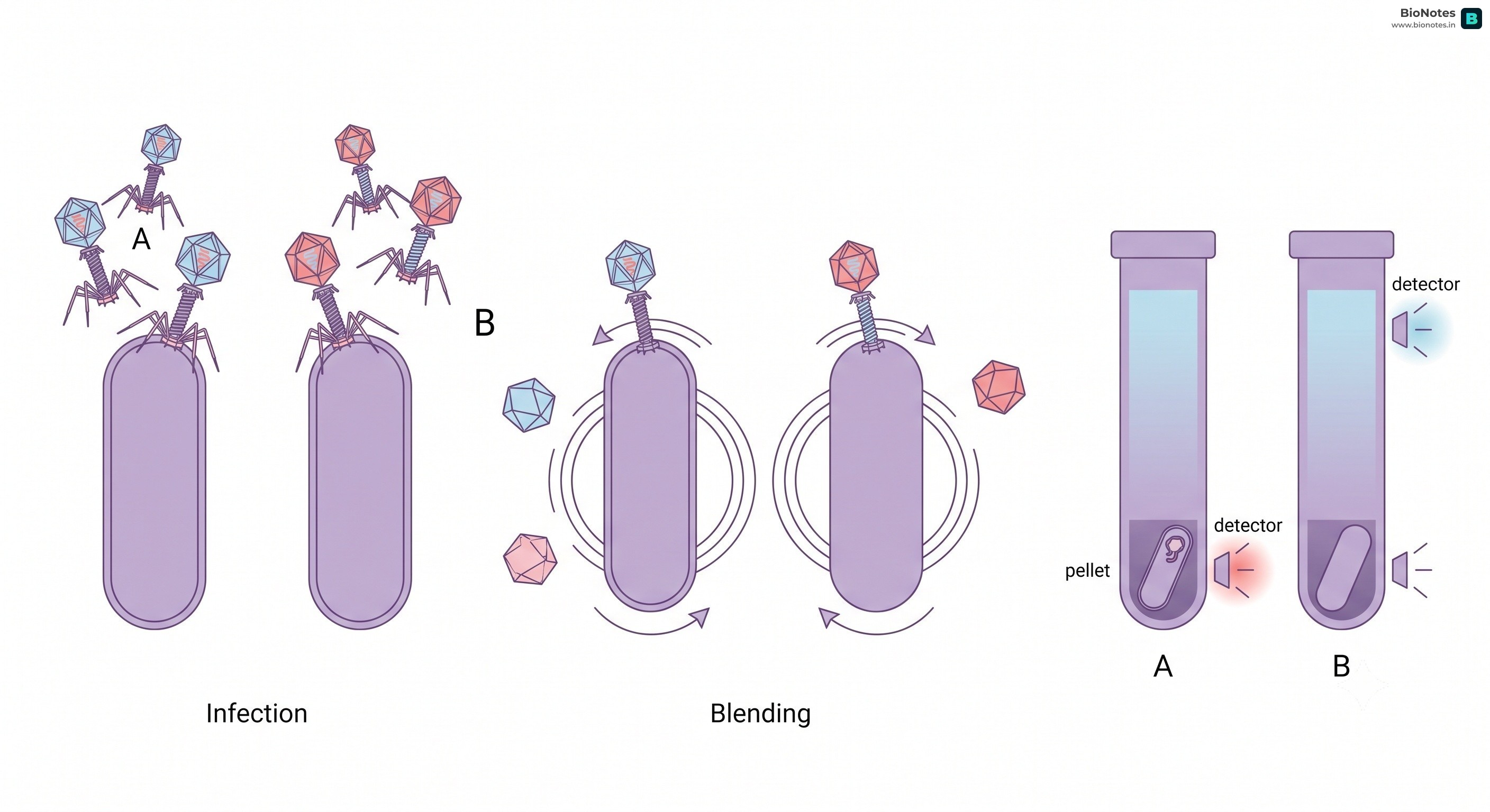
Alfred Hershey and Martha Chase provided unequivocal proof that DNA is the genetic material, using bacteriophages (viruses that infect bacteria).
Experiment:
They grew some bacteriophages on a medium containing radioactive phosphorus (³²P) to label DNA (DNA contains P, protein does not).
They grew others on a medium containing radioactive sulfur (³⁵S) to label proteins (proteins contain S, DNA does not).
These labeled phages were allowed to infect E. coli bacteria.
Blending: The viral coats were agitated off the bacteria in a blender.
Centrifugation: The mixture was centrifuged to separate the viral particles from the bacterial cells.
Observations:
Bacteria infected with ³²P-labeled phages were radioactive, and ³²P was found in the progeny phages.
Bacteria infected with ³⁵S-labeled phages were not radioactive, and ³⁵S remained in the supernatant (viral coats).
Conclusion: DNA, not protein, is the genetic material that is passed from phage to bacteria.
RNA as Genetic Material (Fraenkel-Conrat and Singer Experiment, 1957):
Heinz Fraenkel-Conrat and B. Singer proved that RNA is the genetic material in viruses that do not possess DNA, using two distinct strains of Tobacco Mosaic Virus (TMV): Type A and Type HR (Holmes Ribgrass).
Procedure: They separated the RNA genomes from their protein capsids and reconstituted chimeric hybrid viruses:
Hybrid 1: Type A RNA encapsulated in Type HR protein coat.
Hybrid 2: Type HR RNA encapsulated in Type A protein coat.
Observation: When tobacco plants were inoculated with Hybrid 1, the newly synthesized viral progeny isolated from the infected lesions exclusively possessed Type A RNA and Type A protein coats.
Conclusion: The viral phenotype and progeny characteristics are solely dictated by the RNA molecule, demonstrating unequivocally that RNA serves as the hereditary genetic material in plant and animal RNA viruses (e.g., TMV, QB bacteriophage, Influenza, Polio, and Retroviruses like HIV).
DNA (Deoxyribonucleic Acid): A polymer of deoxyribonucleotides.
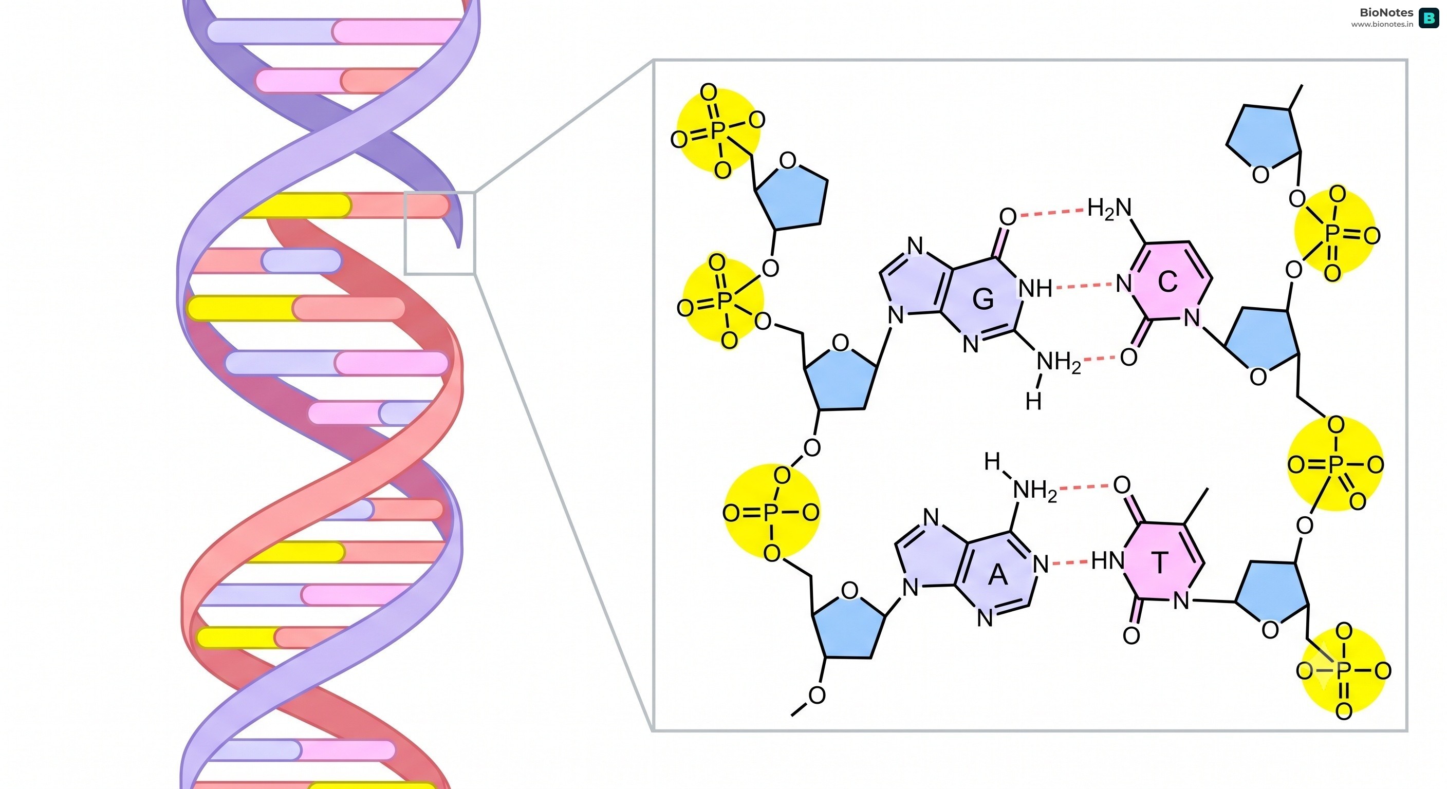
Watson and Crick Model (1953): Based on X-ray diffraction data (Rosalind Franklin and Maurice Wilkins) and Chargaff's rules, James Watson and Francis Crick proposed the double helix model of DNA.
Key Features:
DNA is made of two polynucleotide chains.
The backbones are sugar-phosphate, and the bases project inwards.
The two chains have anti-parallel polarity (one runs 5'→3', the other 3'→5').
The bases in the two strands are paired through hydrogen bonds (A=T, G≡C).
Adenine (A) always pairs with Thymine (T) with two hydrogen bonds.
Guanine (G) always pairs with Cytosine (C) with three hydrogen bonds.
The two chains are coiled in a right-handed helix.
Pitch of the helix is 3.4 nm, with approximately 10 base pairs per turn.
The distance between two base pairs is 0.34 nm.
Did You Know?
The total length of DNA in a single human cell is about 2 meters! If you uncoiled all the DNA in your body and stretched it out, it would reach the Moon and back several times.
Chargaff’s Rule:
Erwin Chargaff (1950) observed that in DNA:
The amount of Adenine (A) is always equal to the amount of Thymine (T) (A=T).
The amount of Guanine (G) is always equal to the amount of Cytosine (C) (G=C).
The ratio of (A+G) to (T+C) is always equal to one.
The ratio of (A+T) to (G+C) varies from species to species.
RNA (Ribonucleic Acid): A polymer of ribonucleotides. Generally single-stranded, but can fold back on itself to form complex 3D structures.
Types of RNA:
mRNA (messenger RNA): Carries genetic information from DNA in the nucleus to the ribosomes in the cytoplasm for protein synthesis. It is linear.
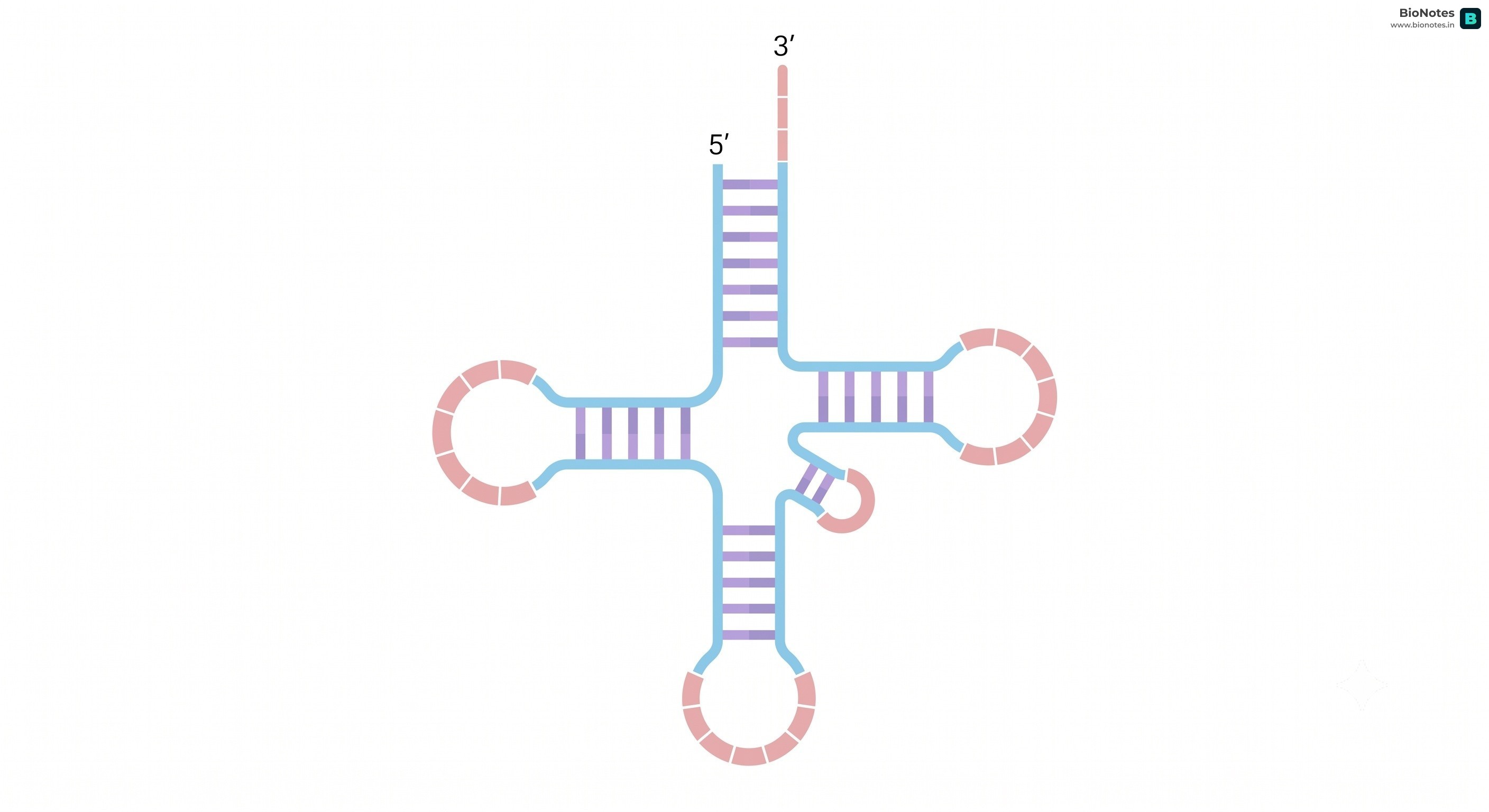
tRNA (transfer RNA): Smallest RNA, acts as an adaptor molecule. It has an anticodon loop (binds to mRNA codon) and an amino acid acceptor end (binds to specific amino acid). It has a clover-leaf like 2D structure and an L-shaped 3D structure.
Interactive Diagram•study Mode
||
www.bionotes.in
rRNA (ribosomal RNA): Most abundant RNA, a structural and catalytic component of ribosomes. It provides the site for protein synthesis.
hnRNA (heterogeneous nuclear RNA): The primary transcript in eukaryotes, contains both introns and exons. It undergoes processing to form mRNA.
snRNA (small nuclear RNA): Involved in splicing of hnRNA.
In Prokaryotes: Cells lack a defined nucleus. The long, negatively charged circular DNA is held in large loops by positively charged non-histone proteins in a distinct cytoplasm region termed the nucleoid.
In Eukaryotes: The total length of DNA in a single diploid human cell is approximately 2.2 meters (6.6×109 bp×0.34×10−9 m/bp), which must be packaged into a microscopic nucleus with a diameter of only ∼10−6 m (10μm). This requires hierarchical compaction:
Histone Proteins: Positively charged, basic proteins rich in the basic amino acid residues Lysine and Arginine. Both amino acids carry positive charges in their side chains, forming tight electrostatic attractions with the negatively charged phosphate groups (PO43−) of DNA.
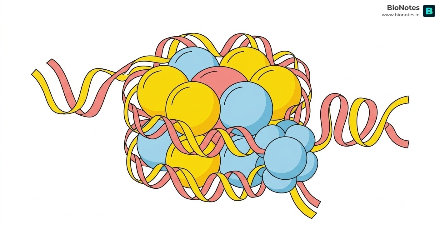
The Histone Octamer: Eight histone molecules assemble to form a core particle (two molecules each of H2A, H2B, H3, and H4).
Nucleosome Structure:
Approximately 200 base pairs (bp) of DNA double helix wrap around the histone octamer in 1.75 left-handed superhelical turns.
H1 Histone (Linker Histone): Placed outside the octamer core, it binds the entry and exit sites of the DNA strand, locking the nucleosome into position.
Adjacent nucleosomes are connected by a segment of linker DNA (∼54 bp), creating a flexible "beads-on-a-string" appearance under electron microscopy (diameter: ∼11 nm).
Higher-Order Chromatin Compaction:
"Beads-on-a-string" chromatin coils into a 30 nm solenoid fibre (6 nucleosomes per turn).
Further looped and folded onto a protein scaffold to form 300 nm chromatin loops, condensed into 700 nm chromatids, and reaches maximum compaction in the 1400 nm metaphase chromosome.
Packaging at higher levels requires an additional set of proteins referred to as Non-Histone Chromosomal (NHC) proteins, which include structural scaffold proteins, topoisomerases, and regulatory transcription factors.
Stains lightly with basic nuclear dyes (e.g., Feulgen, Acetocarmine)
Stains darkly and intensely throughout the cell cycle
Transcriptional State
Transcriptionally active; open for RNA polymerases and transcription factors
Transcriptionally inactive / silent; access to DNA is sterically blocked
Location in Nucleus
Dispersed throughout the central interior of the nucleoplasm
Concentrated along the nuclear periphery (inner nuclear membrane) and chromocenters
Replication Timing
Replicates early in the S phase of the cell cycle
Replicates late in the S phase
Epigenetic Marks
High histone acetylation (loosens histone-DNA contact), low DNA methylation
High DNA methylation and histone methylation (e.g., H3K9me3), low acetylation
Types
Uniform throughout somatic cells
Can be Constitutive (permanently silent, e.g., centromeres, telomeres) or Facultative (inactivated conditionally, e.g., Barr body / inactive X-chromosome)
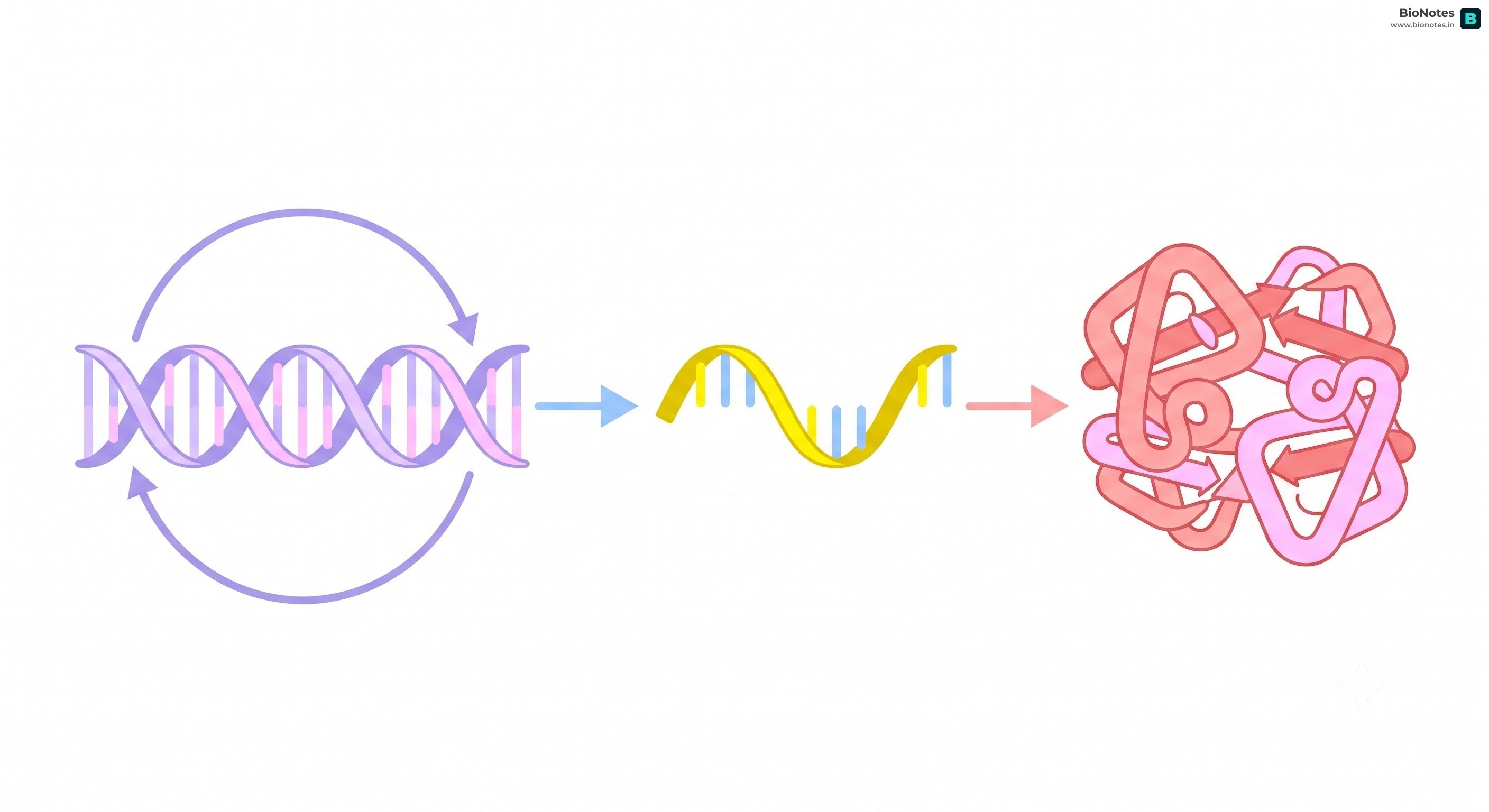
Central Dogma of Molecular Biology: Proposed by Francis Crick.
States that genetic information flows from DNA → RNA → Protein.
Key Concept: The Flow of Information
While the Central Dogma is the standard, remember that Reverse Transcription (RNA → DNA) is a critical exception found in retroviruses like HIV.
Reverse Transcription (Teminism):
Discovered independently by Howard Temin and David Baltimore (1970) in Rous Sarcoma Virus (RSV), earning them the Nobel Prize in 1975.
In retroviruses (e.g., HIV, RSV, oncoviruses), the unidirectional flow of the Central Dogma is reversed:
Viral RNAReverse TranscriptasecDNA (Complementary DNA)→mRNA→Viral Proteins
Catalyzed by the enzyme Reverse Transcriptase (RNA-dependent DNA polymerase), which also possesses RNase H activity (degrades template RNA) and DNA-dependent DNA polymerase activity (synthesizes complementary second DNA strand).
The double-stranded viral DNA integrates into the host genome as a provirus using viral integrase.
DNA Replication: The process by which a DNA molecule makes an exact copy of itself.
Semi-conservative Replication: Proposed by Watson and Crick, and experimentally proven by Meselson and Stahl.
Each new DNA molecule consists of one original (parental) strand and one newly synthesized strand.
Did You Know?
DNA replication is incredibly fast! In E. coli, it happens at a rate of about 2,000 base pairs per second. This means the entire genome is copied in less than 40 minutes.
Meselson and Stahl Experiment (1958):
Interactive Diagram•study Mode
||
www.bionotes.in
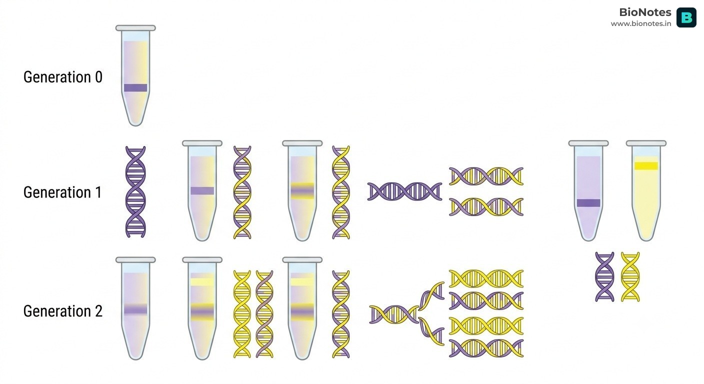
They grew E. coli in a medium containing ¹⁵N (heavy isotope of nitrogen) for several generations, so that ¹⁵N was incorporated into the bacterial DNA.
Then, they transferred the bacteria to a medium containing ¹⁴N (light isotope of nitrogen).
After one generation, the DNA was extracted and centrifuged in a CsCl density gradient. They found an intermediate hybrid band (¹⁵N-¹⁴N).
After two generations, they found two bands: one hybrid (¹⁵N-¹⁴N) and one light (¹⁴N-¹⁴N).
This confirmed the semi-conservative nature of DNA replication.
Enzymes and Proteins Involved in DNA Replication:
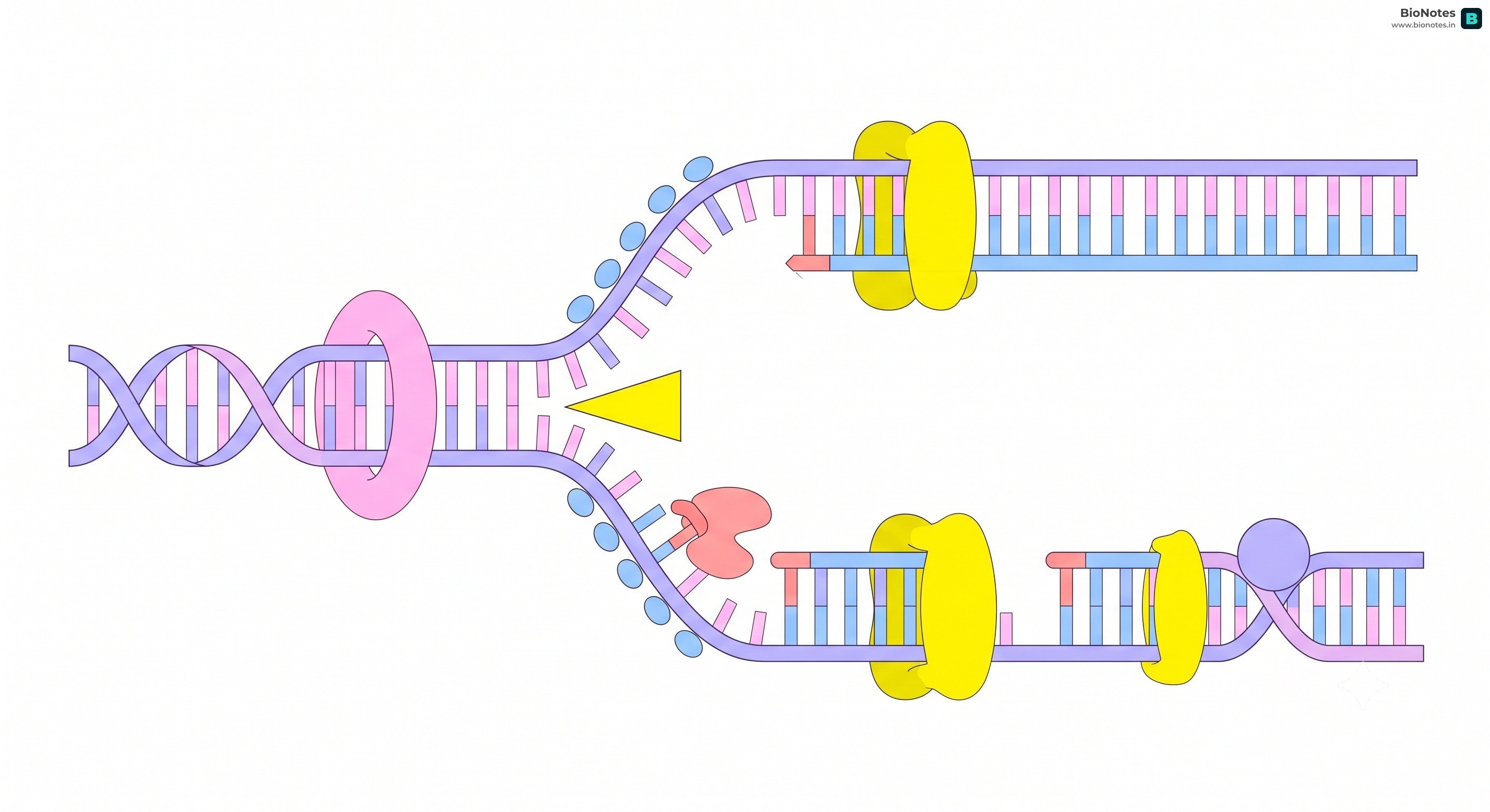
DNA Helicase: Unwinds the DNA double helix at the replication fork by breaking hydrogen bonds between complementary base pairs.
Single-Strand Binding Proteins (SSBs): Bind to and stabilize the exposed single strands of DNA, preventing them from re-annealing (snapping back together) or forming hairpins during replication.
DNA Polymerase: Synthesizes new DNA strands by adding complementary deoxynucleotides. It works exclusively in the 5'→3' direction and requires a free 3'-OH group to initiate synthesis.
In prokaryotes, DNA Pol III is the main replicating enzyme, while DNA Pol I removes RNA primers and fills the gaps (exonuclease activity).
DNA Ligase: Catalyzes the formation of phosphodiester bonds to join the discontinuous Okazaki fragments on the lagging strand.
Primase: Synthesizes a short RNA primer that provides the free 3'-OH group required by DNA polymerase to begin synthesis.
Topoisomerase (DNA Gyrase): Relieves the torsional strain and supercoiling created ahead of the replication fork as the DNA unwinds.
Because the two strands of the parental DNA double helix run anti-parallel, and DNA-dependent DNA polymerase can catalyze polymerization strictly in the 5′→3′ direction, replication proceeds differently on the two template strands:
Parameter
Leading Strand
Lagging Strand
Parental Template Polarity
3′→5′ (towards the replication fork)
5′→3′ (towards the replication fork)
Continuity of Synthesis
Continuous synthesis throughout replication
Discontinuous synthesis in short segments (Okazaki fragments)
Direction Relative to Fork
Synthesized towards the advancing replication fork
Synthesized in short bursts away from the advancing replication fork
Primer Requirement
Requires only a single RNA primer at the replication origin
Requires multiple RNA primers (a separate primer for every Okazaki fragment)
Fragment Size
Continuous, full-length strand
∼1000−2000 nt in prokaryotes; ∼100−200 nt in eukaryotes
DNA Ligase Dependency
Minimal (only at termination / replicon fusion)
Essential; continuously joins adjacent Okazaki fragments by phosphodiester bonds
Synthesis Rate
Faster; unhindered processive elongation
Slightly slower due to repeated cycles of re-priming, primer removal, and fragment ligation
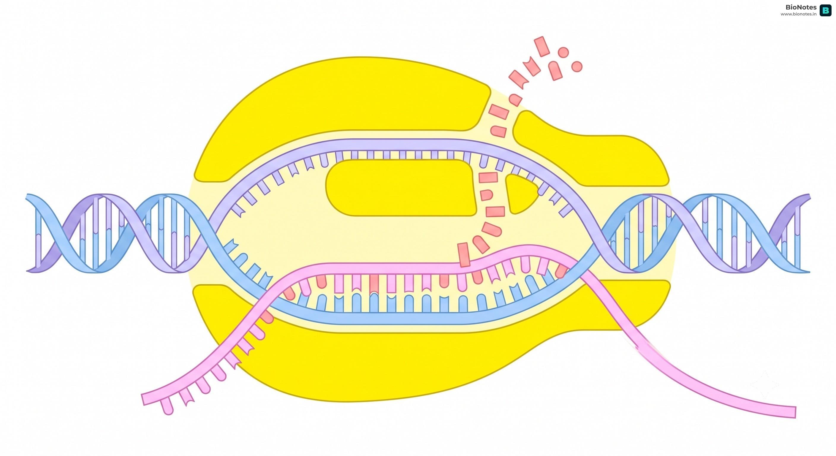
Transcription: The enzymatic process of synthesizing RNA from a DNA template. It is the first step of gene expression where genetic information stored in the nucleotide sequence of DNA is copied into a complementary strand of RNA (mRNA, tRNA, or rRNA).
Fundamental Principles:
Enzyme: Catalyzed by DNA-dependent RNA polymerase.
Rule of Complementarity: Transcription adheres strictly to Watson-Crick base-pairing rules, with one vital distinction: Adenine pairs with Uracil (A=U) instead of Thymine (A=T).
Unidirectional Synthesis: RNA polymerase can only synthesize RNA in the 5′→3′ direction, reading the template strand in the 3′→5′ direction.
Why Both Strands of DNA Are Not Copied During Transcription
In contrast to DNA replication where both parent strands serve as templates, during transcription only one strand of DNA is transcribed. Why?
Coding Ambiguity & Double Genetic Output: If both DNA strands were transcribed, they would produce two RNA molecules with mutually complementary but completely different nucleotide sequences. During translation, these would synthesize two distinct polypeptides with different amino acid sequences. Consequently, one genetic locus would code for two different proteins, leading to utter cellular chaos and corrupted hereditary fidelity.
Double-Stranded RNA (dsRNA) Formation: If two complementary RNA strands were synthesized simultaneously from the same DNA segment, they would readily hybridize through hydrogen bonding to form a double-stranded RNA (dsRNA) molecule. The ribosome cannot bind or translate dsRNA; thus, protein synthesis would be completely abolished, rendering transcription futile.
A transcription unit in double-stranded DNA consists of three essential structural components:
Promoter: A specific non-coding DNA sequence located towards the 5′ end (upstream) of the structural gene (relative to the coding strand). It acts as the binding and recognition platform for RNA polymerase and determines which strand serves as the template.
Structural Gene: The intermediate DNA sequence flanked by the promoter and terminator that encodes the RNA molecule.
Terminator: A sequence located towards the 3′ end (downstream) of the coding strand that marks the end of transcription and triggers the detachment of RNA polymerase and the release of the nascent RNA.
Template Strand vs. Coding Strand:
Template Strand (Antisense / Non-coding strand): Has polarity 3′→5′. Serves as the actual physical template for RNA polymerase. Its sequence is complementary and antiparallel to the newly synthesized RNA.
Coding Strand (Sense / Non-template strand): Has polarity 5′→3′. It does not participate directly in transcription. Its sequence is identical to the synthesized RNA, except that Thymine (T) in DNA is replaced by Uracil (U) in RNA.
Nomenclature Rule: By convention, all directional coordinates and reference positions (such as "upstream", "downstream", promoter at 5′, terminator at 3′) are defined strictly relative to the coding strand.
Feature
Template Strand (Antisense)
Coding Strand (Sense)
Polarity
3′→5′
5′→3′
Role in Transcription
Serves as template for RNA polymerase
Does not act as template
Sequence Relation to RNA
Complementary and antiparallel
Identical (except Thymine in DNA is Uracil in RNA)
Reference Significance
Read by RNA polymerase
Reference for defining upstream (5′) and downstream (3′)
Other Names
Non-coding strand, (−) strand
Non-template strand, (+) strand
Competitive Edge: Cracking the 'Coding Strand' Trap
Board and competitive exam questions frequently test strand polarity:
Question: "If the coding strand sequence is 5′-ATGCATGC-3′, what is the transcribed mRNA sequence?"
Shortcut: You do NOT need to take the complement or invert the direction! The coding strand already has the exact same sequence and polarity as the mRNA. Simply replace T with U:
mRNA=5′-AUGCAUGC-3′
If the question provides the template strand (3′-TACGTACG-5′), then determine the complementary base in the 5′→3′ direction:
mRNA=5′-AUGCAUGC-3′
Cistron: A segment of DNA coding for a single polypeptide chain. It represents the structural gene in molecular terms.
Monocistronic vs. Polycistronic:
Monocistronic Structural Genes: Found primarily in eukaryotes. A single promoter controls transcription of an mRNA that translates into only one polypeptide.
Polycistronic Structural Genes: Found almost exclusively in prokaryotes (bacteria). A single promoter and polycistronic mRNA encode multiple related polypeptides (e.g., the lac operon transcribes an mRNA encoding β-galactosidase, permease, and transacetylase).
Split-Gene Arrangement (Exons and Introns):
Discovered by Phillip Sharp and Richard Roberts (1977, Nobel Prize 1993).
Eukaryotic genes are interrupted (split):
Exons: Coding or expressed sequences that appear in the mature, processed mRNA and code for amino acids.
Introns: Intervening non-coding sequences that do not appear in mature mRNA. They are transcribed into the primary transcript (hnRNA) but are excised during RNA splicing before the mRNA leaves the nucleus.
In bacteria, a single DNA-dependent RNA polymerase transcribes all three major classes of cellular RNA: mRNA, tRNA, and rRNA.
The Holoenzyme Architecture:
Core Enzyme (α2ββ′ω): Catalyzes phosphodiester bond synthesis and elongation, but lacks the ability to recognize and bind specifically to promoter sequences.
Sigma (σ) Factor: An initiation protein that reversibly joins the core enzyme to assemble the RNA Polymerase Holoenzyme (α2ββ′ωσ). It recognizes promoter consensus sequences and unwinds the DNA helix.
The prokaryotic transcription cycle consists of three distinct phases:
Initiation:
The RNA polymerase holoenzyme recognizes and binds to two conserved promoter sequences upstream of the transcription start site (+1):
−35 Sequence:5′-TTGACA-3′ (initial recognition and binding).
−10 Sequence (Pribnow Box):5′-TATAAT-3′ (rich in AT pairs, facilitating strand separation due to 2 hydrogen bonds per pair).
The bound enzyme unwinds approximately 14 base pairs of DNA double helix, transitioning from a "closed promoter complex" to an open promoter complex (transcription bubble).
Synthesis begins with the incorporation of the first ribonucleotide (typically a purine, ATP or GTP).
Elongation:
After an initial oligonucleotide of approximately 8–10 nucleotides is synthesized, the σ factor dissociates from the complex and is recycled.
The core RNA polymerase continues down the template strand in the 3′→5′ direction, synthesizing the RNA strand in the 5′→3′ direction at a rate of 40–50 nucleotides per second.
Ribonucleoside triphosphates (ATP, GTP, CTP, UTP) serve as both substrates and energy sources (rNTP→rNMP+PPi).
A short hybrid RNA-DNA helix (~8–9 base pairs) is maintained within the transient transcription bubble, while the newly synthesized RNA strand is displaced and exits the enzyme.
Termination:
When the core enzyme encounters a terminator sequence, RNA synthesis ceases, the RNA transcript is released, and the enzyme dissociates from DNA. Termination occurs through one of two mechanisms:
Intrinsic (Rho-Independent) Termination:
The terminator sequence contains an inverted repeat rich in G-C base pairs followed by a poly-A sequence on the template strand.
As this region is transcribed, the newly made RNA transcript folds into a stable hairpin (stem-loop) structure held by triple-bonded GC pairs.
Immediately following the hairpin is a run of uracil residues (U6−U8). The hydrogen bonding between rU and dA is exceptionally weak (only 2 H-bonds). The mechanical pull of the hairpin together with the unstable rU:dA hybrid causes RNA polymerase to stall and release the RNA transcript.
Rho (ρ)-Dependent Termination:
The terminator lacks the poly-U stretch.
A hexameric ATP-dependent RNA helicase called Rho (ρ) factor binds to a cytosine-rich, unstructured site on nascent RNA known as the rut (rho utilization) site.
Rho hydrolyzes ATP to translocate along the RNA towards the moving transcription complex. When RNA polymerase pauses at the terminator, Rho catches up, unwinds the RNA-DNA hybrid, and dislodges the transcript and polymerase.
Coupled Transcription and Translation in Prokaryotes
Because prokaryotes lack a nuclear envelope, their genetic material lies freely in the cytoplasm. Furthermore, bacterial mRNA requires no splicing or modifications and is functional immediately upon synthesis.
As a result, translation begins before transcription is even completed. Ribosomes assemble on the free 5′ end of the nascent mRNA and initiate polypeptide synthesis while the 3′ end is still being transcribed by RNA polymerase. Multiple ribosomes translating the same mRNA simultaneously form polyribosomes (polysomes).
In eukaryotic cells, transcription is more complex due to cellular compartmentalization and epigenetic packaging:
Spatial Separation: Transcription and post-transcriptional processing occur exclusively inside the nucleus, whereas translation occurs in the cytoplasm.
Division of Labor — Three Nuclear RNA Polymerases:
Unlike bacteria which use a single RNA polymerase, eukaryotes possess three distinct RNA polymerases in the nucleus, each dedicated to transcribing specific classes of genes:
Enzyme
Cellular Location
Primary Transcripts Synthesized
Sensitivity to α-Amanitin
RNA Polymerase I
Nucleolus
Larger rRNAs: 28S, 18S, and 5.8S rRNA
Insensitive
RNA Polymerase II
Nucleoplasm
hnRNA (heterogeneous nuclear RNA / pre-mRNA), most snRNAs, microRNAs
Highly Sensitive (inhibited at low conc.)
RNA Polymerase III
Nucleoplasm
tRNA, 5S rRNA, and U6 snRNA
Moderately Sensitive (inhibited at high conc.)
Competitive Edge: The 3 Eukaryotic RNA Polymerases
A high-frequency MCQ trap! Remember the mapping:
RNA Pol I:rRNA (28S, 18S, 5.8S — note that 5S rRNA is made by Pol III!).
RNA Pol II:mRNA precursor (hnRNA).
RNA Pol III:tRNA, 5S rRNA, and snRNAs.
Eukaryotic Promoters and Pre-Initiation Complex (PIC):
TATA Box (Goldberg-Hogness Box): Located at −25 to −30 base pairs upstream of the transcription start site. Consensus sequence: 5′-TATAAA-3′.
General Transcription Factors (GTFs): Eukaryotic RNA Polymerase II cannot recognize promoters independently. It requires the sequential assembly of transcription factors:
TFIID (specifically its subunit TBP, TATA-Binding Protein) binds to the TATA box.
TFIIA, TFIIB, TFIIF (which escorts RNA Pol II), TFIIE, and TFIIH assemble to form the Pre-Initiation Complex (PIC).
TFIIH acts as an ATP-dependent helicase to unwind DNA and possesses kinase activity that phosphorylates the C-Terminal Domain (CTD) of RNA Pol II, releasing it from the promoter to commence elongation.
The primary transcript synthesized by RNA Polymerase II is hnRNA (heterogeneous nuclear RNA). It contains non-coding introns, lacks protected termini, and is non-functional. It must undergo three obligatory modifications inside the nucleus to yield mature, translatable mRNA:
5' Capping:
Occurs co-transcriptionally when the nascent RNA transcript is ~20–30 nucleotides long.
An unusual nucleotide, 7-methylguanosine triphosphate (m7Gppp), is added to the 5′ end of hnRNA.
This reaction creates an unusual 5′−5′ triphosphate linkage (unlike the usual 3′−5′ phosphodiester bond).
Functions:
Shields the mRNA from degradation by 5′→3′ exoribonucleases.
Serves as a specific recognition beacon for the small ribosomal subunit (40S) to bind and initiate translation.
Signals nuclear export machinery for transport to the cytoplasm.
3' Polyadenylation (Tailing):
An endonuclease cleaves the pre-mRNA transcript ~10–30 nucleotides downstream of a conserved polyadenylation signal sequence: 5′-AAUAAA-3′.
The enzyme Poly(A) Polymerase (PAP) adds 200–300 adenylate residues to the free 3′-OH end in a template-independent reaction.
Functions:
Protects the 3′ terminus against 3′→5′ exonucleases.
Aids in the export of mature mRNA from the nucleus through nuclear pore complexes.
Stabilizes the transcript and, in synergy with the 5′ cap, promotes translation initiation.
RNA Splicing:
Introns (non-coding intervening sequences) are excised, and exons (coding sequences) are ligated together in a contiguous sequence.
The Spliceosome Machinery:
Splicing is executed by a 60S ribonucleoprotein macromolecular machine called the Spliceosome.
The spliceosome consists of five small nuclear RNAs (snRNAs: U1, U2, U4, U5, and U6) complexed with over 100 proteins to form snRNPs ("snurps" — small nuclear ribonucleoproteins).
Conserved Splice Site Motifs (The GU-AG Rule):
5′ splice site (donor): Invariant GU dinucleotide.
Branch site: Located 20–50 nucleotides upstream of the 3′ splice site; features a conserved internal Adenosine (A).
3′ splice site (acceptor): Invariant AG dinucleotide.
Mechanism (Two-Step Transesterification):
Step 1: The 2′-OH group of the branch-point adenosine performs a nucleophilic attack on the phosphodiester bond at the 5′ splice site (GU). This cleaves the 5′ exon and links the 5′ end of the intron to the branch point adenosine via an unusual 2′−5′ phosphodiester bond, forming a looped structure called a lariat intermediate.
Step 2: The free 3′-OH of the upstream exon attacks the phosphodiester bond at the 3′ splice site (AG). This ligates the two exons together with a standard 3′−5′ phosphodiester bond and releases the intron as a free lariat, which is subsequently linearized and degraded.
Evolutionary Significance & Alternative Splicing:
RNA World Legacy: The catalytic reactions of the spliceosome are carried out by RNA molecules (snRNAs) rather than proteins, acting as ribozymes. This provides evolutionary evidence that RNA served as the primordial biocatalyst in early life.
Alternative Splicing: Eukaryotic pre-mRNAs can undergo alternative splicing, where exons are included or excluded in various combinations. This enables a single gene to encode multiple distinct protein isoforms, explaining how the ~20,000–25,000 human genes can produce over 100,000 unique functional proteins.
Genetic Code: The dictionary of nucleotide base triplets (codons) on mRNA that determines the sequence of amino acids in a polypeptide chain during protein synthesis.
Codon: A sequence of three consecutive ribonucleotides on mRNA that specifies a single amino acid or signals the termination of translation.
Discovering how a 4-letter nucleotide alphabet (A, U, G, C) codes for a 20-letter amino acid alphabet required collaborative insights from physicists, chemists, and biochemists:
George Gamow (1954 - Theoretical Physicist): Postulated that since there are only 4 nitrogenous bases and 20 standard amino acids, a single base (41=4) or a doublet of bases (42=16) would be mathematically insufficient. Therefore, genetic code must be a triplet of bases (43=64 possible codons), providing more than enough permutations to code for 20 amino acids.
Har Gobind Khorana (Nobel Prize 1968): Developed sophisticated chemical methods to synthesize defined RNA homopolymers (e.g., UUUU…) and alternating copolymers (e.g., UGUGUG…), directly linking specific nucleotide sequences to peptide products.
Marshall Nirenberg & J. Heinrich Matthaei (1961): Created a cell-free translation system. When artificially synthesized poly-U (UUUU…) was added to the extract, it catalyzed the synthesis of polyphenylalanine, proving that UUU codes for Phenylalanine. Subsequent trials with poly-C (CCCC…) yielded polyproline, and poly-A (AAAA…) yielded polylysine.
Severo Ochoa (Nobel Prize 1959): Discovered the enzyme polynucleotide phosphorylase (Severo Ochoa enzyme), which polymerizes ribonucleotides into RNA in a template-independent manner with defined enzymatic sequences.
Triplet Nature: Every codon consists of three consecutive nitrogenous bases. 61 codons specify amino acids, and 3 act as stop codons.
Degeneracy: Most amino acids are specified by more than one codon (e.g., Leucine and Arginine each have 6 codons). Degeneracy minimizes the lethal effects of point mutations (silent mutations).
Unambiguous and Specific: A particular codon always codes for one and only one amino acid. For example, UUU codes exclusively for Phenylalanine and never for any other amino acid.
Comma-less (Continuous): The genetic code is read continuously from a fixed start point without any punctuation or pauses between adjacent codons.
Non-overlapping: Consecutive codons are read in succession; each nucleotide is part of only one triplet codon and is never shared between adjacent codons.
Polarity: The code has a definite polarity and is always transcribed and translated in the 5′→3′ direction.
Dual-Purpose Initiator Codon (AUG):
Codes for the amino acid Methionine in eukaryotes (and N-formylmethionine in prokaryotes).
Acts as the universal Start Codon, initiating translation on the ribosome.
(Note: Rarely, in bacteria, GUG or UUG can serve as an alternative initiator codon, but when present at the start position, they still code for fMet).
Universality and Its Exceptions:
The genetic code is universal across almost all living forms (from E. coli to humans, UUU codes for Phenylalanine). This universality is the foundational basis of recombinant DNA technology (allowing human insulin to be expressed in bacteria).
Exceptions to Universality:
Mitochondrial DNA: In human mitochondria, UGA codes for Tryptophan (instead of Stop), AUA codes for Methionine (instead of Isoleucine), and AGA / AGG serve as Stop codons (instead of Arginine).
Ciliated Protozoans (e.g., Paramecium, Tetrahymena):UAA and UAG code for Glutamine instead of signaling Stop.
The Wobble Hypothesis (Francis Crick, 1966):
The pairing between the first two bases of an mRNA codon and the corresponding last two bases of a tRNA anticodon strictly obeys standard Watson-Crick rules (A=U,G≡C).
However, the third base of the codon (at the 3′ end) and the first base of the anticodon (at the 5′ end) has structural flexibility, called "wobble".
Significance: A single tRNA species can recognize multiple degenerate codons differing at the third base. Consequently, cells do not need 61 distinct tRNA species; a set of ∼30−40 tRNAs is fully sufficient to decode all 61 sense codons.
Translation: The biological process in which the sequence of nucleotide triplets (codons) in an mRNA molecule is decoded to synthesize a specific polypeptide chain with a defined amino acid sequence.
The Adaptor Hypothesis (tRNA as the Adaptor):
Francis Crick postulated that amino acids cannot directly read the nucleotide sequence on mRNA because they lack structural and chemical complementarity to nucleic acid bases.
Therefore, an adaptor molecule is required: tRNA (transfer RNA).
Each tRNA features:
An Anticodon Loop containing a triplet base sequence complementary to the mRNA codon.
An Amino Acid Acceptor End at the 3′ terminal (5′-CCA-OH-3′) where the specific amino acid is covalently attached.
Before translation can begin, amino acids must be activated and linked to their cognate tRNAs:
Amino Acid Activation: An amino acid reacts with ATP in the presence of a specific Aminoacyl-tRNA Synthetase enzyme (20 distinct enzymes, one for each amino acid), yielding an enzyme-bound aminoacyl-adenylate intermediate and pyrophosphate (PPi):
Amino Acid+ATPAminoacyl-tRNA SynthetaseAminoacyl-AMP-Enzyme+PPi
tRNA Charging: The activated aminoacyl group is transferred to the 3′-OH of the adenosine residue at the CCA stem of the tRNA, forming a high-energy ester linkage (Aminoacyl-tRNA):
Aminoacyl-AMP-Enzyme+tRNA→Aminoacyl-tRNA (Charged tRNA)+AMP+Enzyme
Energetic Significance: The high-energy ester bond stored in charged tRNA provides the thermodynamic driving force for spontaneous peptide bond synthesis on the ribosome without requiring direct ATP hydrolysis during elongation.
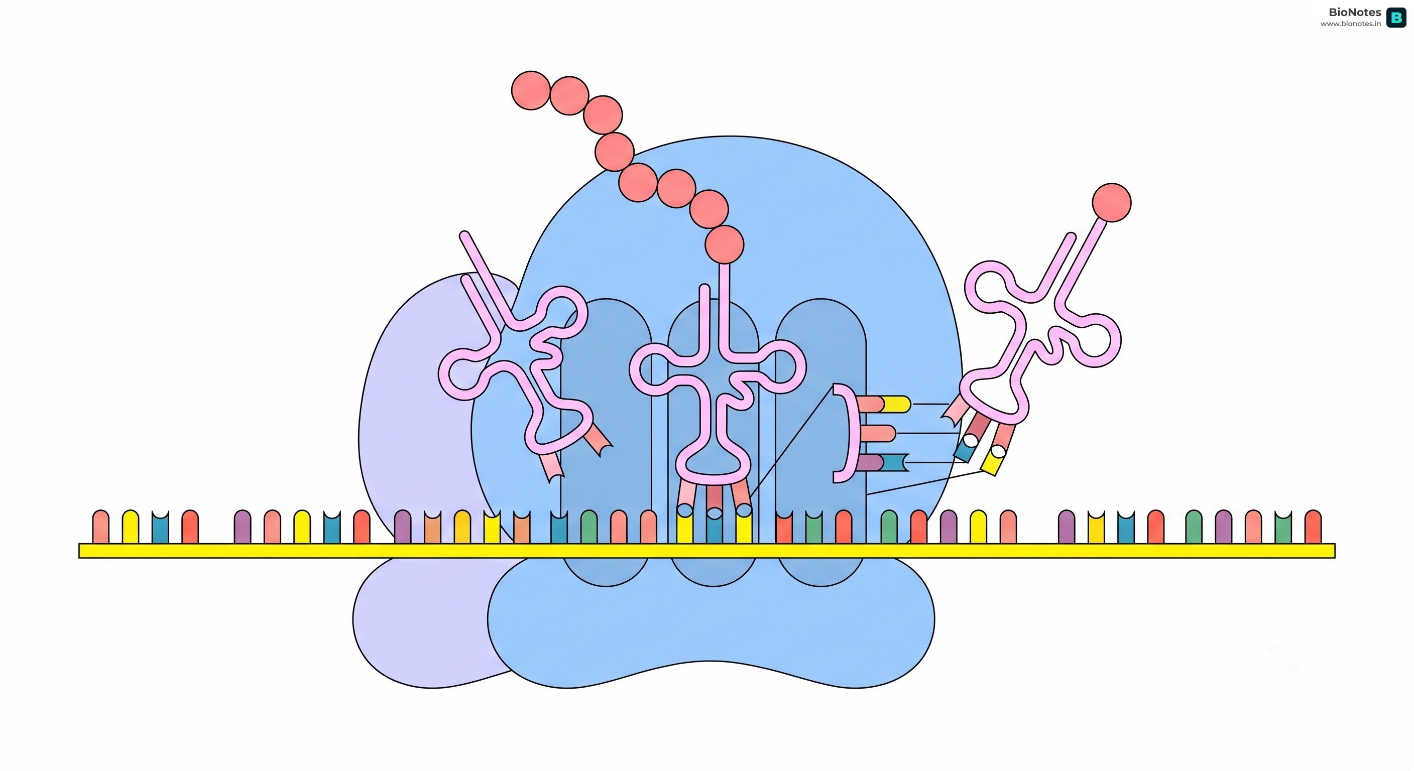
The ribosome is a massive ribonucleoprotein machine consisting of ribosomal RNAs (rRNAs) and approximately 80 proteins:
Subunit Organization:
Prokaryotic Ribosome (70S): Large 50S subunit (contains 23S rRNA and 5S rRNA) + Small 30S subunit (contains 16S rRNA).
Eukaryotic Ribosome (80S): Large 60S subunit (contains 28S rRNA, 5.8S rRNA, and 5S rRNA) + Small 40S subunit (contains 18S rRNA).
Three Functional tRNA Pockets:
A-Site (Aminoacyl / Acceptor site): Binds the incoming aminoacyl-tRNA whose anticodon matches the codon currently positioned in the decoding center.
P-Site (Peptidyl site): Holds the tRNA carrying the growing nascent polypeptide chain. (Crucial Note: The initiator tRNA is the only tRNA that binds directly to the P-site; all subsequent tRNAs enter at the A-site!)
E-Site (Exit site): Holds the deacylated (uncharged) tRNA after peptide bond formation, prior to its ejection back into the cytosol.
The Ribozyme (Peptidyl Transferase):
Peptide bond synthesis between adjacent amino acids is catalyzed by the rRNA of the large subunit acting as a ribozyme (not a protein enzyme!).
In prokaryotes, the catalytic ribozyme is 23S rRNA.
In eukaryotes, the catalytic ribozyme is 28S rRNA.
UTRs (Untranslated Regions): Segments of mRNA that flank the coding region, located at both the 5′ end (upstream of the start codon) and the 3′ end (downstream of the stop codon).
Functions: UTRs do not code for protein, but are essential for efficient translation, mRNA stability, and regulating translation rate.
In Prokaryotes (Shine-Dalgarno Sequence): A conserved purine-rich sequence (5′-AGGAGG-3′) located 8–10 nucleotides upstream of the start codon in the 5′ UTR. It base-pairs with a complementary pyrimidine-rich sequence at the 3′ end of the 16S rRNA in the 30S subunit, aligning the ribosome precisely over the start codon (AUG).
In Eukaryotes (Kozak Sequence): The small 40S subunit binds the 5′ methylguanosine cap and scans downstream along the 5′ UTR until it identifies the start codon within a conserved context: 5′-ACCAUGG-3′ (Kozak consensus sequence).
The small ribosomal subunit binds to mRNA at the initiation site (via the Shine-Dalgarno sequence in prokaryotes or 5′ cap scanning in eukaryotes).
The initiator tRNA carrying N-formylmethionine (fMet-tRNAfMet in bacteria) or Methionine (Met-tRNAiMet in eukaryotes) pairs with the start codon (AUG) directly in the P-site.
Assisted by Initiation Factors (IF1, IF2-GTP, IF3 in prokaryotes; eIFs in eukaryotes).
The large ribosomal subunit docks onto the complex; GTP is hydrolyzed to release initiation factors, assembling the intact 70S/80S initiation complex.
Elongation:
Codon Recognition: The next codon is exposed in the vacant A-site. An elongation factor (EF-Tu bound to GTP in bacteria; eEF1A in eukaryotes) escorts the matching aminoacyl-tRNA to the A-site. Correct base pairing triggers GTP hydrolysis, releasing EF-Tu-GDP.
Peptide Bond Formation: The catalytic 23S rRNA (28S in eukaryotes) in the large subunit cleaves the ester bond between the polypeptide and the tRNA in the P-site, forming a covalent peptide bond between the carboxyl group of the P-site amino acid and the amino group of the A-site amino acid. The growing chain now rests on the tRNA in the A-site.
Translocation: The elongation factor EF-G (translocase) hydrolyzes GTP to ratchet the ribosome three nucleotides (1 codon) forward along mRNA in the 5′→3′ direction.
The uncharged tRNA shifts from the P-site to the E-site and is discharged.
The peptidyl-tRNA shifts from the A-site to the P-site.
The A-site becomes vacant, exposing the next codon for the next incoming tRNA.
Termination:
Elongation continues until one of the three stop codons (UAA, UAG, UGA) enters the A-site.
Stop codons are not recognized by any tRNA. Instead, protein Release Factors (RF1 recognizes UAA/UAG; RF2 recognizes UAA/UGA; RF3-GTP promotes release in prokaryotes; eRF1 recognizes all three codons in eukaryotes) bind the stop codon in the A-site.
The peptidyl transferase center adds a water molecule instead of an amino acid to the peptidyl-tRNA, cleaving the ester bond and releasing the newly synthesized polypeptide chain.
Ribosome Recycling Factors (RRF) and EF-G trigger the complete dissociation of the ribosomal subunits, releasing mRNA and tRNAs for reuse.
Both transcription and translation represent the core engine of the Central Dogma, converting static genomic blueprints into functional enzymatic and structural machinery:
Parameter
Transcription
Translation
Core Definition
Enzymatic copying of a DNA sequence into an RNA molecule (mRNA, tRNA, rRNA)
Decoding of nucleotide codons on an mRNA molecule into a polypeptide chain
Central Dogma Flow
DNA → RNA (First step of gene expression)
RNA → Protein (Second step of gene expression)
Cellular Site (Prokaryotes)
Cytoplasm
Cytoplasm (coupled simultaneously with transcription)
Cellular Site (Eukaryotes)
Nucleus (nucleoplasm / nucleolus)
Cytoplasm (cytosol and Rough Endoplasmic Reticulum)
Template Molecule
Template strand of double-stranded DNA (3′→5′)
Single-stranded messenger RNA (mRNA) (5′→3′)
Direction Template is Read
Read in the 3′→5′ direction
Read in the 5′→3′ direction
Direction of Product Synthesis
Synthesized 5′→3′
Synthesized from N-terminus (amino end) → C-terminus (carboxyl end)
Gene Expression: The process by which information from a gene is used in the synthesis of a functional gene product (protein or RNA).
Regulation of Gene Expression: In prokaryotes, gene expression is predominantly regulated at the transcriptional initiation level.
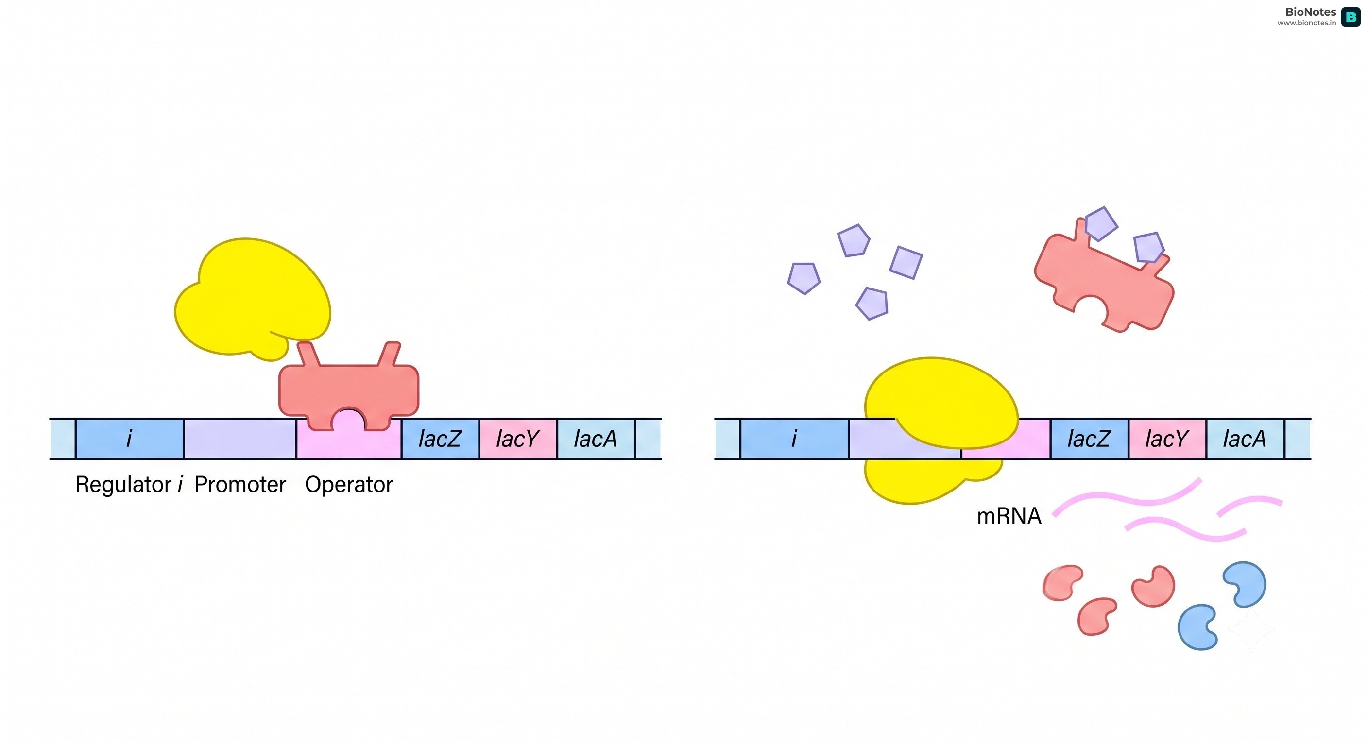
Lac Operon (in E. coli): Elucidated by François Jacob and Jacques Monod (1961, Nobel Prize 1965). It is an inducible operon governing the transport and catabolism of lactose.
Components of the Operon:
Promoter (P): The DNA binding site for RNA polymerase.
Operator (O): The regulatory DNA sequence adjacent to the promoter that binds the repressor protein.
Structural Genes: Transcribed as a single polycistronic mRNA:
lacZ: Encodes β-galactosidase, which hydrolyzes lactose into glucose and galactose (and isomerizes lactose into allolactose).
lacY: Encodes β-galactoside permease, which increases the cell membrane's permeability to lactose.
lacA: Encodes β-galactoside transacetylase, which transfers an acetyl group from acetyl-CoA to β-galactosides for detoxification.
Regulator Gene (i gene): Codes constitutively for the Lac Repressor protein (a homotetramer). Note: "i" stands for inhibitor, not inducer!
When Lactose is Absent (Switched OFF):
The active repressor protein synthesized by the i gene binds firmly to the operator region (O).
This physically blocks RNA polymerase from transcribing the structural genes.
Operon is switched OFF (basal transcription remains at a negligible trace level).
When Lactose is Present (Switched ON):
A small amount of lactose enters the cell via basal permease and is converted into allolactose by β-galactosidase.
Allolactose acts as the inducer. It binds to an allosteric site on the repressor protein, causing a conformational change.
The altered repressor loses affinity for the operator and detaches from the DNA.
RNA polymerase can now transcribe the structural genes (lacZ, lacY, lacA), switching the operon ON.
Diauxic Growth: When E. coli is cultured in a medium containing both glucose and lactose, it selectively metabolizes glucose first until it is completely exhausted, pauses in a brief diauxic lag phase, and then induces the lac operon to utilize lactose.
Mediated by cAMP (cyclic AMP) and CAP (Catabolite Activator Protein), also called CRP (cAMP Receptor Protein).
High Glucose: Cellular cAMP levels are low; CAP remains inactive and cannot bind DNA. Transcription of the lac operon is weak even if lactose is present.
Low / No Glucose: Cellular cAMP levels rise sharply; cAMP binds CAP, forming an active cAMP-CAP complex.
The cAMP-CAP complex binds to a specific CAP recognition site upstream of the promoter, bending the DNA and recruiting RNA polymerase with high affinity. This produces a 50-fold increase in transcription.
Summary Rule: The lac operon is maximally expressed only when lactose is present AND glucose is absent.
Human Genome Project (HGP): An international mega-scientific collaborative initiative launched in 1990 and completed in 2003 (with the complex sequence of Chromosome 1 finished in May 2006). It was coordinated by the U.S. Department of Energy (DOE) and the National Institutes of Health (NIH), with core global partnerships in the UK (Wellcome Trust), Japan, France, Germany, and China.
Why HGP Was Designated a "Mega-Project":
Enormous Scale: The human haploid genome comprises approximately 3×10⁹ base pairs.
Financial Investment: At an estimated cost of US 3perbasepair,theprojectedbudgetwasroughly∗∗US9 billion**.
Data Magnitude: If sequence data were printed in books of 1,000 pages each, with each page containing 1,000 letters, it would take 3,300 volumes just to record a single human genome!
Birth of Bioinformatics: Managing this immense volume of raw data necessitated the inception of Bioinformatics—a hybrid discipline fusing biology, computer science, and data analytics.
The project adopted two distinct methodological strategies:
Expressed Sequence Tags (ESTs): Focused strictly on identifying and sequencing only those genes that are transcribed into functional RNA (the coding transcriptome).
Blind Sequence Annotation (Whole Genome Shotgun Sequencing): The unbiased approach of shearing and sequencing the entire genome (both coding and non-coding regions) into overlapping fragments, which were subsequently assembled into contigs using supercomputers.
Cloning Vectors & Host Systems: Fragmented human DNA was cloned into bacterial (E. coli) and yeast (Saccharomyces cerevisiae) hosts using specialized, high-capacity vectors:
BAC (Bacterial Artificial Chromosomes)
YAC (Yeast Artificial Chromosomes)
Sequencing Technology: Performed using automated DNA sequencers based on the dideoxy chain-termination technique pioneered by Frederick Sanger (two-time Nobel Laureate, who also determined the first amino acid sequence of a protein, insulin).
The completion of the HGP unveiled fundamental molecular truths that revolutionized genetics:
Total Genome Size: The human genome contains 3,164.7 million (3.16×10⁹) base pairs.
Gene Size Variation: An average human gene consists of ∼3,000 bases, but sizes vary dramatically. The largest known human gene is Dystrophin (2.4×10⁶ base pairs / 2.4 Mb on the X-chromosome), while one of the smallest is the SRY/TDF gene (14 bp).
Low Total Gene Count: The total number of genes is estimated at 20,000−25,000—vastly fewer than previous scientific predictions of 80,000−100,000.
High Sequence Homology: Almost 99.9% of nucleotide bases are identical in all human beings! Only a tiny fraction (0.1%, equivalent to ∼3 million bp) accounts for all phenotypic and disease variations among individuals.
Unknown Gene Functions: The biological functions of over 50% of discovered genes remain completely unknown.
Low Protein-Coding Proportion:Less than 2% of the human genome encodes functional proteins; over 98% is non-coding.
Repetitive Sequences Abundance: Repetitive sequences make up very large portions of the genome. While they have no direct coding function, they provide vital clues regarding chromosome architecture, chromosomal dynamics, and evolutionary mutations.
Gene Density Disparities:Chromosome 1 has the most genes (2,968), whereas the Y chromosome has the fewest genes (231).
Single Nucleotide Polymorphisms (SNPs): Scientists have mapped approximately 1.4 million locations where single-base DNA differences occur in the human population. These "snips" serve as essential genomic markers for diagnosing hereditary disease susceptibility, customizing pharmacogenomics, and tracing human genealogical lineages.
DNA Fingerprinting (DNA Profiling): A forensic technique used to distinguish and identify individuals based on unique variations in their DNA sequences.
Pioneering Scientists:
Developed internationally by Sir Alec Jeffreys (1984) at the University of Leicester.
Pioneered in India by Dr. Lalji Singh ("Father of Indian DNA Fingerprinting", who developed the indigenous Bkm probe from the banded krait) and Dr. V.K. Kashyap.
Exploiting the 0.1% Divergence: Because 99.9% of human DNA is identical, sequencing the entire genome for forensic identification is impractical. DNA fingerprinting specifically targets the highly variable, non-coding 0.1% of the genome.
Density Gradient Centrifugation (Bulk vs. Satellite DNA):
When genomic DNA is sheared and subjected to Cesium Chloride (CsCl) density gradient ultracentrifugation, it separates into distinct bands based on buoyant density:
Major Bulk Peak: Represents the majority of genomic DNA (contains coding genes and unique sequences).
Classification of Satellite DNA: Based on base composition (A-T rich vs. G-C rich), length of the repeat segment, and total number of repeating units:
Micro-satellites (STRs - Short Tandem Repeats): Very short repeat units of 1 to 6 base pairs (e.g., (CA)n).
Mini-satellites (VNTRs - Variable Number Tandem Repeats): Repeat units of 10 to 100 base pairs, flanked by conserved restriction enzyme cleavage sites. VNTRs belong to a class of satellite DNA called minisatellites.
DNA Polymorphism as the Basis of Identification:
Because satellite DNA does not code for any protein, mutations that arise within these repetitive sequences do not affect an individual's reproductive fitness or phenotype. Consequently, mutations accumulate undisturbed from generation to generation.
An inheritable DNA alteration that occurs in a human population at a frequency greater than 0.01 (1%) is defined as a DNA Polymorphism.
Satellite DNA shows an extraordinarily high degree of polymorphism. The number of tandem repeats at a given chromosomal locus varies widely from person to person.
Mendelian Inheritance: A child inherits 50% of their VNTR alleles from the mother and 50% from the father.
Tissue Invariance: The VNTR fingerprint is completely identical across every cell and tissue of an individual's body (blood, skin, hair follicle, semen, saliva, bone) and remains immutable throughout life. Identical (monozygotic) twins share the exact same DNA fingerprint.
The classic Alec Jeffreys method involves six sequential laboratory steps:
Isolation of DNA: High-molecular-weight genomic DNA is extracted and purified from nucleated biological specimens (blood, semen stains, buccal swabs, hair follicles).
Restriction Digestion: The extracted DNA is digested into fragments using specific restriction endonucleases (e.g., EcoRI, HinfI) that cut outside the VNTR repeat arrays.
Agarose Gel Electrophoresis: The resulting DNA fragments are loaded into an agarose gel and separated according to molecular size under an electric field (smaller fragments migrate faster towards the positive anode).
Alkaline Denaturation: The gel is soaked in an alkaline solution (NaOH) to denature the double-stranded DNA fragments into single strands.
Southern Blotting: The separated single-stranded DNA fragments are transferred from the fragile gel onto a durable synthetic nylon or nitrocellulose membrane via capillary action.
Hybridization with Labeled Probe: The membrane is incubated with single-stranded, radioactively labeled (32P) or fluorescent VNTR probes that selectively bind (hybridize) to their complementary tandem repeat sequences on the membrane.
Autoradiography: The membrane is washed to remove unbound probes and exposed to an X-ray film. Radioactive probes expose the film, producing characteristic dark bands at specific positions. This banding pattern represents the unique DNA fingerprint (resembling a supermarket barcode).
Modern Forensic Upgrade: PCR-Based STR Profiling
Classic VNTR Southern blotting required microgram quantities of high-molecular-weight, undegraded DNA. In modern forensic practice, it has been largely superseded by PCR-amplified Short Tandem Repeats (STRs). Because PCR amplifies minute quantities of DNA exponentially, modern labs can generate complete, courtroom-admissible genetic profiles from picogram traces (such as the root of a single stray hair, trace saliva on a postage stamp, or epithelial skin cells left on a touched surface).
Forensic Investigation: Unequivocally matches biological evidence found at crime scenes (blood, semen, hair, tissue) to suspects or exonerates the wrongfully accused.
Paternity and Maternity Disputes: Conclusively determines biological parentage by comparing child bands against maternal and paternal fingerprints (every band in the child's fingerprint must match either the mother or the father).
Disaster Victim Identification (DVI): Positively identifies victims of mass casualties, wars, plane crashes, and natural disasters from skeletal remains.
Evolutionary Biology & Anthropology: Traces human migratory patterns, genetic lineages, and historical demographic bottlenecks.
Wildlife Conservation: Detects poaching, illegal wildlife trafficking, and establishes genetic diversity in captive breeding programs for endangered species.
Ethical, Legal, and Social Concerns:
Genetic privacy and data protection rights (risks of national DNA database misuse).
Genetic discrimination in health insurance, employment, or marriage.
Chain of custody and contamination risks in forensic sample collection.
 BioNotes