All living organisms are composed of organic and inorganic carbon-based chemicals known as biomolecules. To determine the chemical composition of a living cell, analytical fractionation is performed using trichloroacetic acid.
Homogenization: Fresh living tissue (e.g., plant leaf, liver tissue, or microbial paste) is thoroughly ground in Trichloroacetic acid (Cl3C-COOH) using a mortar and pestle to produce a thick slurry.
Filtration: The slurry is filtered through a double layer of cheesecloth or cotton gauze, yielding two distinct fractions:
Analytical Fraction
Alternative Term
Molecular Weight Range
Molecular Nature
Primary Chemical Constituents
Acid-Soluble Pool
Filtrate
18−800 Da
Biomicromolecules (monomeric units & inorganic ions)
Why do Lipids appear in the Acid-Insoluble Retentate despite having a molecular weight <800 Da?
Lipids are not polymers and strictly have a molecular weight not exceeding 800 Da. By weight, they are micromolecules. However:
Lipids are major components of cellular membranes (plasma membrane, nuclear envelope, endoplasmic reticulum, mitochondrial membranes).
When living tissue is disrupted by mechanical grinding, cellular membranes fragment and spontaneously seal into water-insoluble micelles and vesicles.
These hydrophobic vesicle aggregates fail to dissolve in the polar aqueous trichloroacetic acid medium and are physically trapped on the filter cloth.
Hence, lipids appear in the acid-insoluble retentate solely due to their structural association and hydrophobicity, not because they are true polymers!
Elemental analysis shows that living organisms contain the same basic elements found in the Earth's crust, but in dramatically different relative abundances:
Carbon, Hydrogen, Oxygen, and Nitrogen are significantly enriched in living organisms compared to the Earth's crust.
Silicon (Si) is abundant in the Earth's crust (≈27.7%) but is negligible in living tissue (<0.01%).
Carbohydrates are polyhydroxy aldehydes or polyhydroxy ketones, or organic substances that yield such compounds upon acid hydrolysis. Their general empirical formula is Cn(H2O)n.
The absolute configuration is designated D- or L- based on the spatial orientation of the hydroxyl (−OH) group attached to the penultimate chiral carbon (C-5 in glucose), referenced against D-glyceraldehyde.
If the −OH on C-5 points to the right in a Fischer projection, it is a D-sugar.
If the −OH on C-5 points to the left, it is an L-sugar.
Biological Rule: Naturally occurring sugars in animal and plant metabolism are overwhelmingly D-isomers (e.g., D-glucose, D-fructose, D-ribose), whereas amino acids in proteins are almost exclusively L-isomers.
D-Glucose and L-Glucose are enantiomers (non-superimposable mirror images) with identical physical properties except for the direction in which they rotate plane-polarized light.
In aqueous solution, less than 0.02% of glucose exists in the open-chain aldehyde form. The molecule spontaneously undergoes an intramolecular nucleophilic addition:
The −OH on C-5 attacks the carbonyl carbon at C-1.
This cyclization forms a 6-membered heterocyclic hemiacetal ring termed a pyranose ring (glucopyranose).
Cyclization transforms the planar, achiral C-1 carbonyl carbon into a new asymmetric chiral center termed the Anomeric Carbon.
Depending on the spatial orientation of the newly generated −OH at C-1:
α-D-Glucopyranose: The C-1 −OH points downward in the Haworth projection (trans to the C-6 −CH2OH group).
β-D-Glucopyranose: The C-1 −OH points upward in the Haworth projection (cis to the C-6 −CH2OH group).
Mutarotation is the spontaneous, time-dependent change in optical rotation observed when a freshly prepared solution of an optically pure carbohydrate (α or β anomer) is allowed to stand, until an equilibrium value is reached.
When crystals of pure α-D-glucose are dissolved in water, the initial specific optical rotation is +112.2∘.
Over time, the hemiacetal ring spontaneously opens to reform the free aldehyde open-chain intermediate (<0.02%), which then re-closes to form either α or β anomers.
When crystals of pure β-D-glucose are dissolved in water, the initial specific optical rotation is +18.7∘.
Both solutions gradually converge to the exact same equilibrium optical rotation of +52.7∘.
Thermodynamic Basis for β-Anomer Dominance (64% vs. 36%):
In the chair conformation (4C1) of β-D-glucopyranose, all five bulky substituents (−OH at C-1, C-2, C-3, C-4 and −CH2OH at C-5) occupy the sterically favored equatorial positions.
In α-D-glucopyranose, the anomeric −OH at C-1 is forced into an axial position, causing destabilizing 1,3-diaxial steric interactions.
Consequently, β-D-glucopyranose is thermodynamically more stable (ΔG∘<0) and constitutes ≈64% of the equilibrium mixture.
Disaccharides consist of two monosaccharide units joined by a covalent glycosidic bond (formed by condensation with the loss of one water molecule).
Disaccharide
Constituent Monomers
Glycosidic Bond Type
Reducing Property
Occurrence & Biological Function
Maltose
α-D-Glucose+α-D-Glucose
α(1→4)
Reducing (free C-1 hemiacetal on 2nd glucose)
Starch breakdown product in germinating barley
Lactose
β-D-Galactose+β-D-Glucose
β(1→4)
Reducing (free C-1 hemiacetal on glucose)
Principal sugar in mammalian milk (4−7%)
Sucrose
α-D-Glucose+β-D-Fructose
α(1↔2)β
Non-reducing (both anomeric carbons linked)
Table sugar, primary transport sugar in phloem
Trehalose
α-D-Glucose+α-D-Glucose
α(1↔1)α
Non-reducing (both anomeric carbons linked)
Hemolymph of insects, cryptobiotic desiccation protectant
The Phenomenon of Invert Sugar (Inversion of Cane Sugar)
Pure Sucrose is dextrorotatory ([α]D=+66.5∘).
When hydrolyzed by dilute acid or the enzyme invertase (sucrase), it cleaves into an equimolar mixture of:
D-Glucose: Dextrorotatory ([α]D=+52.7∘).
D-Fructose: Strongly levorotatory ([α]D=−92.4∘).
Calculating net optical rotation of the equimolar hydrolysate:
Net [α]D=2+52.7∘+(−92.4∘)=−19.85∘(Levorotatory)
Because the sign of optical rotation inverts from dextro (+) to levo (−), the resulting equimolar mixture is called Invert Sugar, and the reaction is called the Inversion of Sucrose (found in natural honey).
Amylose (15−20%): Unbranched linear chain of D-glucose units linked by α(1→4) bonds. Adopts a helical secondary structure that can trap molecular iodine (I2) inside its helical coils, producing an intense deep blue color.
Amylopectin (80−85%): Branched chain. Linear segments have α(1→4) links; branch points occur every 24−30 residues via α(1→6) linkages. Produces a reddish-violet color with iodine.
Glycogen ("Animal Starch"):
Major storage polysaccharide in animal liver and skeletal muscle.
Structure is similar to amylopectin but much more extensively branched (branch points occur every 8−12 glucose residues).
Gives a reddish-brown color with iodine.
Cellulose:
Most abundant organic biomolecule in the biosphere.
Linear unbranched polymer of D-glucose linked by β(1→4) glycosidic bonds.
In cellulose, each alternate glucose residue is rotated 180∘ relative to its neighbor. Chains remain strictly linear and form extensive intra- and inter-chain hydrogen bonds, assembling into rigid, insoluble microfibrils.
No Helical Coils: Because it lacks helical secondary structure, cellulose cannot trap I2 and gives no blue color with iodine!
Chitin:
Second most abundant organic polymer on Earth.
Homopolymer of N-acetyl-β-D-glucosamine (NAG) joined by β(1→4) linkages.
Found in the rigid exoskeletons of arthropods (insects, crustaceans) and the cell walls of fungi.
Inulin:
Polymer of fructose (β−2,1 links) ending in a terminal glucose residue.
Stored in dahlias and chicory roots.
Filtered freely by the renal glomerulus without being reabsorbed or secreted; used clinically to measure Glomerular Filtration Rate (GFR).
An α-amino acid is a substituted methane consisting of four distinct groups attached to a central asymmetric carbon atom, designated the α-Carbon (Cα):
An amino group (−NH2)
A carboxyl group (−COOH)
A hydrogen atom (−H)
A distinctive variable side chain (−R group)
        H
        │
   H₂N ─ Cα ─ COOH
        │
        R
Chirality & Optical Activity:
All 19 standard amino acids are chiral and optically active.
Glycine (R=−H) is the sole exception: its Cα is bonded to two identical hydrogen atoms, making it achiral (optically inactive).
Absolute Configuration:
Ribosomally synthesized proteins incorporate exclusively L-amino acids (the −NH2 group is situated on the left in the standard Fischer projection).
- Tyrosine & Tryptophan strongly absorb ultraviolet light at 280 nm, used universally in spectrophotometric protein quantification. - Tyrosine has a phenolic −OH group (pKR=10.1).
- Cysteine: Possesses a reactive sulfhydryl (−SH) group; oxidation of two cysteines forms a covalent disulfide bond (−S-S−), yielding Cystine. - Serine & Threonine: Contain aliphatic −OH groups (targets for protein kinase phosphorylation).
Acidic (Negatively Charged at pH 7.0)
Carboxylate side chains
Aspartate (Asp, D) Glutamate (Glu, E)
- Contain extra carboxyl groups (pKR≈3.9−4.2). - Form electrostatic salt bridges with basic amino acids.
- Arginine: Contains a positively charged guanidinium group (pKR≈12.5, strongest base). - Lysine: Possesses an ϵ-amino group (pKR≈10.5). - Histidine: Possesses an imidazole ring (pKR≈6.0); acts as the primary physiological buffer in hemoglobin and blood around cellular pH 7.4.
Amino acids are amphoteric substances containing both acidic (−COOH) and basic (−NH2) ionizable groups. In aqueous solution, an intramolecular acid-base proton transfer occurs: the −COOH donates a proton to the −NH2 group, forming a dipolar ion called a Zwitterion (German: hybrid ion).
          IONIZATION EQUILIBRIA OF AN AMINO ACID ACROSS pH

          Low pH (Acidic)            Neutral pH (pI)           High pH (Alkaline)
             pH < pKa1                  pH = pI                    pH > pKa2

                H                          H                           H
                │                          │                           │
         ⁺H₃N ─ C ─ COOH            ⁺H₃N ─ C ─ COO⁻              H₂N ─ C ─ COO⁻
                │                          │                           │
                R                          R                           R

         Cationic Form                 Zwitterion                 Anionic Form
        (Net Charge = +1)          (Net Charge = 0)            (Net Charge = -1)
       Migrates to Cathode (-)     No Electrophoretic         Migrates to Anode (+)
                                       Migration
The Isoelectric Point (pI) is the precise pH at which an amino acid possesses zero net electrical charge (equal positive and negative charges) and therefore does not migrate in an applied electrical field.
For Neutral Amino Acids (Monoamino-monocarboxylic, e.g., Alanine, Glycine):pI=2pKa1(α-COOH)+pKa2(α-NH3+)Example (Alanine):pKa1=2.34, pKa2=9.69⟹pI=22.34+9.69=6.015.
For Acidic Amino Acids (Containing an ionizing carboxyl R-group, e.g., Aspartate, Glutamate):pI=2pKa1(α-COOH)+pKR(R-COOH)Averaging the two lowest pKa values because the neutral zwitterionic form exists between these two transitions.Example (Aspartate):pKa1=2.1, pKR=3.9, pKa2=9.8⟹pI=22.1+3.9=3.0.
For Basic Amino Acids (Containing an ionizing nitrogenous R-group, e.g., Lysine, Arginine):pI=2pKR(R-NH3+)+pKa2(α-NH3+)Averaging the two highest pKa values.Example (Lysine):pKa1=2.2, pKa2=9.0, pKR=10.5⟹pI=29.0+10.5=9.75.
Physical Significance of pI in Laboratory & Medicine
At its isoelectric point (pH=pI):
Net electrostatic charge on the protein is zero.
Electrostatic repulsion between adjacent protein molecules is minimized.
Protein solubility reaches an absolute minimum, causing proteins to aggregate and precipitate out of solution (Isoelectric Precipitation).
Used extensively in protein purification techniques like Isoelectric Focusing (IEF) and 2D Gel Electrophoresis.
Peptides and proteins are formed when the α-carboxyl group of one amino acid condenses with the α-amino group of another amino acid with the elimination of one molecule of water, yielding a peptide bond (−CO-NH−):
                       RESONANCE OF THE PEPTIDE BOND

             O                         O⁻
             ║                         │
      ─ Cα ─ C ─ N ─ Cα ─   <=====>   ─ Cα ─ C ═ N⁺ ─ Cα ─
                 │                               │
                 H                               H

         Single Bond State                 Double Bond State
           (~60% weight)                     (~40% weight)
Partial Double-Bond Character: Due to resonance delocalization of the non-bonding electron pair on the nitrogen atom into the carbonyl oxygen π-orbital, the peptide C-N bond possesses approximately 40% double-bond character.
Bond Length: The peptide C-N bond length is 1.32A˚, which is distinctly shorter than a normal single C-N bond (1.47A˚) and longer than a pure double C=N bond (1.27A˚).
Planar Geometry: Free rotation around the C-N bond is completely prevented. The six atoms constituting the peptide unit (Cα1, C, O, N, H, and Cα2) lie strictly within a single rigid plane (the amide plane).
Trans Conformation: Nearly all peptide bonds in native proteins adopt the trans configuration (dihedral angle ω≈180∘) rather than the cis configuration (ω≈0∘). The trans geometry minimizes steric clash between the R-groups of neighboring Cα atoms.
The linear sequence of amino acids linked by covalent peptide bonds, numbered from the N-terminus (free amino group) to the C-terminus (free carboxyl group).
Anfinsen's Dogma (Nobel Prize 1972): Christian Anfinsen demonstrated using ribonuclease A that the primary amino acid sequence contains all the necessary physical instructions required to direct the spontaneous folding of a protein into its native 3D conformation.
Secondary Structure (2∘):
Local regular conformational folding of the polypeptide backbone, stabilized exclusively by hydrogen bonds between the backbone carbonyl oxygen (>C=O) and backbone amide hydrogen (−N-H):
α-Helix: Right-handed spiral. 3.6 amino acid residues per complete helical turn; pitch is 0.54 nm (5.4A˚). Every backbone carbonyl oxygen at residue n forms a hydrogen bond with the amide hydrogen at residue n+4. Proline disrupts this geometry because its cyclic ring lacks a backbone amide hydrogen and induces a severe steric kink (helix breaker).
β-Pleated Sheet: Polypeptide chains extended in a zigzag conformation aligned side-by-side. Hydrogen bonds form laterally between adjacent chains. Can run antiparallel (adjacent chains run in opposite N→C directions; linear, highly stable H-bonds) or parallel (chains run in the same direction; skewed H-bonds).
Tertiary Structure (3∘):
The complete, overall three-dimensional folding of a single polypeptide chain into a compact globular or fibrous conformation.
Stabilizing Forces:
Hydrophobic Interactions: The primary thermodynamic driving force (hydrophobic collapse). Nonpolar side chains (Leu, Ile, Val, Phe) bury inside the interior core away from water, while polar residues face the external solvent.
Hydrogen Bonds: Between polar R-groups.
Ionic Bonds / Salt Bridges: Electrostatic attraction between oppositely charged groups (e.g., −COO− of Asp and −NH3+ of Lys).
Disulfide Bridges: Covalent −S-S− bonds between Cysteine residues; provides extraordinary thermal and chemical stability (e.g., insulin, antibodies, keratin).
Van der Waals Forces: Weak induced dipolar attractions between nonpolar atoms packed closely together.
Quaternary Structure (4∘):
The spatial arrangement and assembly of two or more independent polypeptide chains (subunits or protomers) into a functional multi-subunit oligomer.
Classic Example:Adult Human Hemoglobin (HbA) is a heterotetramer consisting of two identical α-subunits (141 amino acids each) and two identical β-subunits (146 amino acids each) assembled into an α2β2 architecture.
Although the peptide bond itself is rigid and planar, rotation is permissible around the two single covalent bonds connecting each α-carbon atom to the rest of the polypeptide backbone.
ϕ (Phi): The angle of rotation around the single bond between the backbone nitrogen and the α-carbon (N−Cα).
ψ (Psi): The angle of rotation around the single bond between the α-carbon and the carbonyl carbon (Cα−C).
ω (Omega): The torsion angle around the peptide bond itself (C−N), fixed at ≈180∘ for trans.
                   POLYPEPTIDE BACKBONE TORSION ANGLES

                   H   R       O       H   R
                   │   │       ║       │   │
             ── N ─ Cα ─ C ── N ─ Cα ─ C ──
                │        │    │        │
                └───┬────┘    └───┬────┘
                  φ (Phi)       ψ (Psi)
               (N - Cα bond) (Cα - C bond)
In 1963, eminent Indian biophysicist Gopalasamudram Narayana Ramachandran (G.N. Ramachandran), along with C. Ramakrishnan and V. Sasisekharan at the University of Madras, plotted ψ against ϕ on a Cartesian coordinate plane spanning from −180∘ to +180∘.
They discovered that because atoms behave as hard spheres with defined Van der Waals radii, steric clashes between non-bonded backbone atoms and side-chain R-groups render approximately 75% of all theoretical (ϕ,ψ) angle combinations physically impossible (forbidden)!
Left-handed α-helix (αL):ϕ≈+57∘,ψ≈+47∘. Standard L-amino acids can rarely adopt this conformation due to steric overlap between the side-chain Cβ and carbonyl oxygen; only glycine residues comfortably occupy this region in native structures.
Quadrant IV (ϕ>0,ψ<0, Bottom-Right):
Sterically Disallowed: Severe Van der Waals overlaps between the side-chain atoms and backbone carbonyl oxygen render this zone forbidden for all amino acids (except glycine).
Structural Validation:
In structural biology (X-ray crystallography, Cryo-EM), software tools like PROCHECK plot every residue of a solved protein on a Ramachandran diagram. A high-resolution, biologically valid structure must have >90% of all non-glycine residues in the core favored regions.
For over 50 years, resolving the "Protein Folding Problem"—predicting the complex three-dimensional folded atomic structure of a protein purely from its one-dimensional primary amino acid sequence—was one of the greatest grand challenges in natural science (Levinthal's paradox estimated 10100 possible conformations).
In 2020, Google DeepMind unveiled AlphaFold 2, which solved this challenge with atomic-level accuracy, culminating in the 2024 Nobel Prize in Chemistry (awarded to Demis Hassabis and John Jumper of Google DeepMind, and David Baker of the University of Washington for computational protein design).
A per-residue confidence metric on a scale from 0 to 100:
pLDDT>90 (Very High Confidence - Dark Blue): High-accuracy backbone and side-chain orientations; comparable to high-resolution X-ray crystallographic coordinates (<1.5A˚ RMSD).
50<pLDDT≤70 (Low Confidence - Yellow): Flexible loops, unstructured surface linkers, or mobile domains.
pLDDT<50 (Very Low Confidence - Orange): Strong bioinformatic marker for Intrinsically Disordered Proteins (IDPs) or Intrinsically Disordered Regions (IDRs). These segments do not adopt a stable 3D conformation in isolation and only fold upon binding physiological partners.
PAE (Predicted Aligned Error):
A 2D matrix estimating the positional error (in Angstroms, A˚) of residue x when the predicted structure is aligned against the true structure at residue y.
Critical for determining whether two distinct protein domains maintain a rigid, fixed spatial relationship relative to one another, or whether they are flexibly hinged.
Lipids are a diverse group of nonpolar, water-insoluble biomolecules that are readily soluble in nonpolar organic solvents (chloroform, ether, benzene).
Fatty acids are organic carboxylic acids with long, unbranched hydrocarbon chains (4 to 36 carbons) terminating in a carboxyl group (−COOH).
Saturated Fatty Acids:
Contain no double bonds (C-C single bonds only).
Chains pack tightly via maximum Van der Waals contacts, resulting in high melting points (solid at room temperature; termed fats).
Palmitic acid:16 carbons (CH3(CH2)14COOH, melting point 63∘C).
Stearic acid:18 carbons (CH3(CH2)16COOH, melting point 69∘C).
Unsaturated Fatty Acids:
Contain one or more double bonds (C=C), naturally occurring almost exclusively in the cis-configuration.
Cis double bonds introduce a rigid 30∘ kink into the hydrocarbon chain, preventing tight packing and lowering the melting point (liquid at room temperature; termed oils).
Simple Lipids: Esters of fatty acids with various alcohols.
Triglycerides (Triacylglycerols / Neutral Fats): Formed by the esterification of one molecule of glycerol (1,2,3-trihydroxypropane) with three fatty acid molecules via three ester bonds.
Compound / Complex Lipids: Esters of fatty acids containing additional chemical functional groups.
Phospholipids: Consist of glycerol, two fatty acids, a phosphate group, and a polar head group.
Classic Example:Lecithin (Phosphatidylcholine). Contains a polar, hydrophilic choline-phosphate head and two nonpolar, hydrophobic fatty acyl tails. This amphipathic structure is the absolute structural prerequisite for the spontaneous assembly of the lipid bilayer of biological membranes.
Derived Lipids: Formed by the hydrolysis of simple and compound lipids, or synthesized from isoprene units.
Steroids: Characterized by a core four-ring carbon skeleton consisting of three cyclohexane rings and one cyclopentane ring fused together: the cyclopentanoperhydrophenanthrene (CPPP) nucleus.
Cholesterol: The principal animal sterol. It modulates eukaryotic membrane fluidity, preventing membrane crystallization at low temperatures and excessive permeability at high temperatures. It serves as the obligatory metabolic precursor for bile salts, vitamin D, and all steroid hormones (cortisol, aldosterone, testosterone, estradiol, progesterone).
Nucleic acids (Deoxyribonucleic Acid, DNA, and Ribonucleic Acid, RNA) are linear, unbranched biopolymers composed of monomeric units termed nucleotides. They function as the primary repository, replication template, and transcriptional expression machinery of genetic information in all cellular life.
Nitrogenous Bases: Planar, aromatic, heterocyclic nitrogen-containing ring structures:
Purines (Bicyclic, Nine-Membered Double Ring): Formed by a six-membered pyrimidine ring fused to a five-membered imidazole ring with nitrogen atoms at positions 1,3,7, and 9.
Adenine (A):6-aminopurine.
Guanine (G):2-amino-6-oxypurine.
Pyrimidines (Monocyclic, Six-Membered Single Ring): Single heterocyclic ring with nitrogen atoms at positions 1 and 3.
Cytosine (C):4-amino-2-oxypyrimidine (present in both DNA and RNA).
Thymine (T):5-methyluracil / 2,4-dioxo-5-methylpyrimidine (exclusive to DNA). The methyl group at C-5 significantly enhances thermal and photochemical stability against UV-induced pyrimidine dimer formation.
Uracil (U):2,4-dioxypyrimidine (exclusive to RNA).
Pentose Sugars (Five-Carbon Monosaccharides):
β-D-Ribose (in RNA): Contains hydroxyl (−OH) groups at both the C-2′ and C-3′ positions.
β-D-2′-Deoxyribose (in DNA): Lacks an oxygen atom at the C-2′ position (possesses a −H atom instead of −OH).
Why is DNA chemically more stable than RNA?
The absence of the 2′−OH group in deoxyribose makes DNA significantly more resistant to spontaneous alkaline hydrolysis. In RNA, the adjacent 2′−OH acts as an internal nucleophile that readily attacks the 3′−5′ phosphodiester linkage under physiological or alkaline conditions, causing rapid strand cleavage. Hence, evolution selected DNA as the stable repository of hereditary information.
Phosphate Group:
Derived from orthophosphoric acid (H3PO4).
Esterified to the C-5′ hydroxyl of the pentose sugar.
Because phosphoric acid has a low pKa, the phosphate groups carry a net negative charge at physiological pH 7.4, imparting a strong polyanionic negative charge to the entire nucleic acid backbone (which enables tight electrostatic binding to basic, positively charged histone octamers in eukaryotic chromatin).
N-Glycosidic Bond: Covalent linkage formed between the C-1′ carbon of the pentose sugar and the N-9 position of a purine or the N-1 position of a pyrimidine, with the elimination of a water molecule.
Phosphoester Bond: Covalent ester linkage formed between the C-5′ hydroxyl of the pentose and a phosphate group.
3′−5′ Phosphodiester Bond: The fundamental repeating covalent linkage that polymerizes individual nucleotides into a continuous polynucleotide chain. The phosphate group forms a bridge connecting the C-3′ hydroxyl of one pentose sugar to the C-5′ hydroxyl of the adjacent pentose sugar.
Chain Polarity: Every polynucleotide chain has an intrinsic directionality:
5′-End: Bears a free phosphate group attached to the C-5′ of the terminal pentose.
3′-End: Bears a free hydroxyl (−OH) group attached to the C-3′ of the terminal pentose.
High-Energy Phosphoanhydride Bonds: In nucleotide triphosphates like ATP and GTP, the α-phosphate is attached to the ribose via a phosphoester bond, whereas the β- and γ-phosphates are linked to each other via phosphoanhydride bonds, which release large amounts of Gibbs free energy (ΔG∘′≈−30.5 kJ/mol or −7.3 kcal/mol) upon hydrolysis.
In 1953, James Watson and Francis Crick deduced the three-dimensional secondary structure of DNA, building on the critical X-ray crystallographic diffraction pattern (Photo 51) obtained by Rosalind Franklin and Maurice Wilkins, along with the chemical base-ratio data of Erwin Chargaff.
Right-Handed Helical Polarity: Two polynucleotide chains wound coaxially around a central symmetry axis in a right-handed helix.
Antiparallel Alignment: The two strands run in opposite directions: one strand has 5′→3′ polarity, and the complementary strand has 3′→5′ polarity.
Core vs. Exterior Organization:
The hydrophilic sugar-phosphate backbones are located on the exterior of the helix, exposed to the surrounding aqueous environment.
The hydrophobic nitrogenous bases project into the interior core, oriented nearly perpendicular (88.5∘) to the central helical axis.
Complementary Base Pairing:
Adenine always pairs specifically with Thymine via two hydrogen bonds (A = T).
Guanine always pairs specifically with Cytosine via three hydrogen bonds (G≡C).
A purine always pairs with a pyrimidine, maintaining an invariant, uniform helical diameter of 2.0 nm (20A˚) across the entire molecule.
Because of the three hydrogen bonds, G-C pairs are thermodynamically more stable than A-T pairs. Genomes or DNA fragments with high GC content possess a significantly higher melting temperature (Tm).
Dimensions & Spatial Helical Parameters:
Helical Pitch (p): The axial length of one complete 360∘ helical turn is 3.4 nm (34A˚).
Base Pairs per Turn: Each turn comprises approximately 10 base pairs (10.4−10.5 bp in aqueous solution).
Distance Between Adjacent Base Pairs (h): The axial rise per base pair is 0.34 nm (3.4A˚).
Angle of Rotation: Each successive base pair rotates by 36∘ relative to the preceding pair (360∘/10 bp).
Major and Minor Grooves:
Because the glycosidic bonds do not project at diametrically opposite angles (180∘) from the sugar rings, the double helix features alternating depressions along its surface:
Major Groove (2.2 nm wide, deep): Exposes distinct hydrogen-bond donors, acceptors, and methyl groups that permit sequence-specific recognition by DNA-binding proteins (transcription factors, restriction endonucleases, zinc fingers).
Minor Groove (1.2 nm wide, shallow): Less information-rich; bound by non-sequence-specific architectural proteins like histones.
Thermodynamic Stabilization Forces:
Hydrogen Bonding: Contributes to specificity of base pairing.
Base Stacking Interactions (π−π stacking): Flat aromatic rings of neighboring base pairs stack directly atop one another in the anhydrous interior, maximizing Van der Waals contacts and hydrophobic interactions. Base stacking provides the predominant thermodynamic stability of the double helix, exceeding the energetic contribution of hydrogen bonds.
In 1950, Austrian-American biochemist Erwin Chargaff analyzed the quantitative nitrogenous base compositions of DNA extracted from diverse organisms and formulated empirical rules that apply strictly to double-stranded DNA (dsDNA):
Purine to Pyrimidine Equivalence: The total molar concentration of purines equals the total molar concentration of pyrimidines:
[A]+[G]=[T]+[C]⟹[T]+[C][A]+[G]=1.0
Equimolar Base Pairs:
The amount of Adenine is always equal to Thymine: [A]=[T]⟹[T][A]=1.0.
The amount of Guanine is always equal to Cytosine: [G]=[C]⟹[C][G]=1.0.
The Base Ratio is Species-Specific:
The ratio of [G]+[C][A]+[T] is not necessarily equal to 1.0.
It is constant for a given biological species, serving as a characteristic taxonomic signature. Organisms with G+CA+T>1 are classified as AT-type (e.g., humans ≈1.52), while those with G+CA+T<1 are classified as GC-type (e.g., Mycobacterium tuberculosis≈0.42).
Exam Warning: When do Chargaff's Rules NOT Apply?
Chargaff's rules apply strictly to double-stranded DNA. They do NOT hold true for:
Single-stranded DNA (ssDNA): e.g., bacteriophage ϕX174 and M13. If a sample has [A]=[T] or [G]=[C], it is diagnostic proof of single-stranded genetic material!
DNA is structurally polymorphic and can adopt alternative helical conformations depending on hydration state, sequence composition, and salt concentration:
Structural Parameter
B-DNA (Canonical Form)
A-DNA (Dehydrated Form)
Z-DNA (Left-Handed Form)
Helical Sense
Right-handed
Right-handed
Left-handed
Occurrence / Conditions
Standard physiological aqueous state (>92% relative humidity)
Low hydration (75% humidity); found in RNA-DNA hybrids and dsRNA
Alternating purine-pyrimidine tracts (e.g., poly(dG-dC)); high salt or cytosine methylation
Diameter of Helix
2.0 nm (20A˚)
2.3 nm (23A˚, broader)
1.8 nm (18A˚, most slender)
Base Pairs per Helical Turn
10 bp (average 10.4−10.5)
11 bp
12 bp (6 repeating dimers)
Helical Pitch (p)
3.4 nm (34A˚)
2.8 nm (28A˚)
4.5 nm (45A˚)
Rise per Base Pair (h)
0.34 nm (3.4A˚)
0.26 nm (2.6A˚)
0.37 nm (3.7A˚)
Base Tilt to Helical Axis
Perpendicular (88.5∘)
Tilted by 20∘
Tilted by 7∘
Sugar Pucker Conformation
C-2′ endo
C-3′ endo
Alternating: C-2′ endo (for pyrimidines) and C-3′ endo (for purines)
Unlike DNA, cellular RNA typically exists as a single-stranded polynucleotide (ssRNA), allowing it to fold back on itself via intramolecular complementary base pairing (A=U,G≡C, and non-canonical G-U wobble pairs) to form intricate secondary structures (hairpin loops, stem-loops, pseudoknots) and catalytic tertiary folds.
                              CELLULAR RNA CLASSES
                                       │
        ┌──────────────────────────────┼──────────────────────────────┐
        ▼                              ▼                              ▼
  Messenger RNA (mRNA)          Ribosomal RNA (rRNA)           Transfer RNA (tRNA)
  - 3 - 5% of total RNA         - ~80% of total RNA            - 10 - 15% of total RNA
  - Linear informational copy   - Structural & Catalytic       - Adaptor molecule
  - Short half-life             - Peptidyl transferase ribozyme - Cloverleaf (2°) / Inverted-L (3°)
Messenger RNA (mRNA, 3−5% of total cellular RNA):
Transcribes genetic instructions from genomic DNA in the nucleus and directs polypeptide synthesis at ribosomes.
Possesses a 5′-untranslated region (5′-UTR), a protein-coding sequence initiated by the start codon AUG, and a 3′-UTR.
In eukaryotes, processing adds a 5′-methylguanosine cap (m7Gppp) and a 3′-polyadenylated tail (poly-A).
Ribosomal RNA (rRNA, ≈80% of total cellular RNA):
The most abundant and metabolically stable class of RNA.
Forms the structural framework and catalytic core of ribosomes:
Prokaryotes (70S Ribosome): Contains 16S rRNA in the 30S small subunit, and 23S rRNA + 5S rRNA in the 50S large subunit. The 23S rRNA is a true ribozyme that catalyzes peptide bond synthesis.
Eukaryotes (80S Ribosome): Contains 18S rRNA in the 40S subunit, and 28S, 5.8S, and 5S rRNA in the 60S subunit.
Transfer RNA (tRNA / Soluble RNA / Adaptor Molecule, 10−15% of total cellular RNA):
Decodes the nucleotide triplets of mRNA into corresponding amino acids during translation.
Secondary Structure (Robert Holley, 1965): Resembles a Cloverleaf, consisting of four distinct arms:
Acceptor Stem: Unpaired 7-base pair stem ending in the conserved single-stranded sequence 5′−CCA−3′ at the 3′-terminus, where the specific amino acid is covalently attached via an ester bond to the 3′−OH of adenosine.
Anticodon Arm: Contains a 7-nucleotide loop bearing the Anticodon triplet that base-pairs antiparallel to the complementary mRNA codon.
D-Arm (Dihydrouridine Arm): Contains modified dihydrouridine; recognized by aminoacyl-tRNA synthetase.
TψC-Arm (Ribothymidine-Pseudouridine-Cytidine Arm): Binds the ribosome surface during protein synthesis.
Variable Arm: Differentiates tRNA classes by length.
Tertiary Structure (Alexander Rich & Aaron Klug, 1974): The cloverleaf folds through tertiary hydrogen bonds into a compact, three-dimensional Inverted L-shape, positioning the amino acid acceptor site at one extreme tip and the anticodon triplet at the opposite tip (≈7.0 nm apart).
Small Non-Coding Regulatory RNAs:
Small Nuclear RNA (snRNA): Combines with proteins to form Small Nuclear Ribonucleoproteins (snRNPs or "snurps") that assemble the eukaryotic spliceosome to excise introns from pre-mRNA.
MicroRNA (miRNA) & Small Interfering RNA (siRNA):21−25 nucleotide regulatory RNAs that mediate post-transcriptional gene silencing via the RNA interference (RNAi) pathway.
Enzymes are specialized biological catalysts that accelerate the rate of metabolic chemical reactions without being permanently altered or consumed in the process.
Ribozymes: Non-Protein Biological Catalysts
While nearly all enzymes are globular proteins, ribozymes are catalytic RNA molecules. Discovered by Thomas Cech and Sidney Altman (Nobel Prize in Chemistry 1989), examples include the 23S rRNA peptidyl transferase of bacterial ribosomes (catalyzing peptide bond formation) and Ribonuclease P.
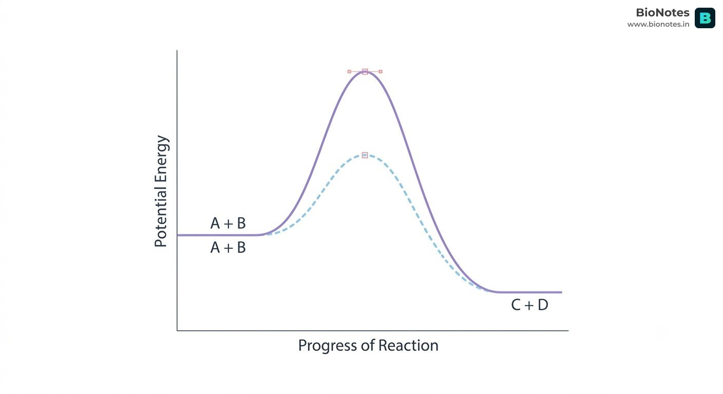
Chemical reactions require an initial threshold of energy, the Activation Energy (Ea), to reach an unstable, high-energy transition state (X‡) before converting into products.
Interactive Diagram•study Mode
||
www.bionotes.in
Catalytic Strategy: An enzyme does not change the overall thermodynamic equilibrium of a reaction, nor does it alter the standard free energy change (ΔG).
Instead, the enzyme binds the substrate at its catalytic active site, stabilizing the transition state through complementary non-covalent interactions and dramatically lowering the activation energy barrier (Ea).
Catalytic Models:
Lock-and-Key Model (Emil Fischer, 1894): Proposes a rigid, static active site geometrically pre-shaped to fit the substrate.
Induced-Fit Model (Daniel Koshland, 1958): The active site is flexible; binding of the substrate induces a conformational rearrangement in the enzyme to achieve optimum catalytic alignment.
The International Union of Biochemistry and Molecular Biology (IUBMB) classifies enzymes into six major classes based on the specific type of reaction catalyzed:
Class Code
Class Name
General Reaction Type Catalyzed
Canonical Examples
EC 1
Oxidoreductases
Oxidation-reduction reactions (transfer of electrons or hydride ions: Ared+Box⇌Aox+Bred)
                           THE MICHAELIS-MENTEN EQUATION

                                      Vmax · [S]
                                V = ─────────────
                                      Km + [S]
Substrate Concentration ([S]):
At low [S], reaction rate V is directly proportional to [S] (first-order kinetics).
At high [S], all enzyme active sites become saturated with substrate, and the rate reaches a plateau designated Vmax (Maximum Velocity) (zero-order kinetics).
Km (Michaelis Constant): The substrate concentration at which the reaction velocity is exactly half of its maximum (21Vmax).
Affinity Indicator:Km is an inverse measure of enzyme-substrate affinity. A low Km value indicates high affinity (the enzyme reaches 21Vmax at very low substrate concentrations).
Temperature:
Velocity rises with temperature due to increased kinetic energy (Q10≈2).
Reaches an Optimum Temperature (typically 37∘C−40∘C for human enzymes).
Above the optimum, thermal vibration ruptures weak tertiary hydrogen and ionic bonds, causing irreversible denaturation and loss of catalytic activity.
pH:
Each enzyme has an Optimum pH (e.g., Pepsin ≈pH 1.5−2.0, Salivary Amylase ≈pH 6.8, Trypsin ≈pH 8.0).
Extreme pH changes alter the ionization states of acidic (−COO−) and basic (−NH3+) amino acids within the active site, disrupting catalytic turnover.
Competitive Inhibition: Inhibitor competes at the active site. Can be completely overcome by flooding the system with excess substrate.
Vmax is UNCHANGED.
Km INCREASES (affinity appears decreased).
Non-Competitive Inhibition: Inhibitor binds to an allosteric regulatory site, distorting active site geometry. Adding more substrate cannot rescue activity.
Vmax DECREASES.
Km is UNCHANGED (the uninhibited active sites retain normal affinity).
Uncompetitive Inhibition: Inhibitor binds only to the enzyme-substrate ([ES]) complex.
Both Vmax and Km DECREASE.
Inhibition Type
Binding Site on Enzyme
Effect on Vmax
Effect on Km
Classic Pharmacological & Biological Examples
Competitive
Active Site (competes directly with substrate)
Unchanged
Increases
1. Malonate inhibits succinate dehydrogenase (competing with succinate in Krebs cycle). 2. Sulfa drugs compete with para-aminobenzoic acid (PABA) for bacterial folic acid synthesis. 3. Statin drugs (e.g., Lovastatin) inhibit HMG-CoA reductase for cholesterol management.
Non-Competitive
Allosteric Site (site other than active site)
Decreases
Unchanged
1. Cyanide (CN−) binds the iron-copper center of cytochrome c oxidase, halting mitochondrial electron transport. 2. Heavy metal toxicity (Pb2+,Hg2+,Ag+) reacting with active site −SH groups.
Feedback (Allosteric) Inhibition
Regulatory Allosteric Site
Decreases
Varies
The end-product of a metabolic pathway acts as an allosteric inhibitor of the first committed pathway step (e.g., Isoleucine allosterically inhibits threonine deaminase; ATP inhibits phosphofructokinase-1).
Many enzymes require non-protein chemical components called co-factors to become catalytically active.
An inactive, purely protein enzyme is called an Apoenzyme.
The catalytically active complex (apoenzyme + cofactor) is termed a Holoenzyme:
Apoenzyme (Protein)+Co-factor (Non-protein)=Holoenzyme (Active Enzyme)
ENZYME CO-FACTORS │ ┌──────────────────────────┴──────────────────────────┐ ▼ ▼ Organic Co-factors Inorganic Co-factors │ │ ┌────┴───────────────────────────┐ ▼ ▼ ▼ Metal IonsProsthetic Groups Co-enzymes (Coordination bridges)- Tightly / Covalently bound - Transiently bound - Zn²⁺ (Carbonic Anhydrase)- Permanent association - Derived from vitamins - Mg²⁺ (Hexokinase, DNA Pol)- E.g., Haem in Peroxidase - E.g., NAD⁺ (Niacin) - Fe²⁺/Fe³⁺ (Cytochromes)
Prosthetic Groups: Organic molecules that are tightly and permanently (often covalently) bound to the apoenzyme.
Example:Haem is the prosthetic group tightly integrated into the catalytic active sites of peroxidase and catalase (which catalyze the breakdown of toxic hydrogen peroxide into water and oxygen).
Co-enzymes: Organic molecules that are transiently and loosely associated with the apoenzyme during catalytic turnover.
Most co-enzymes are metabolic derivatives of water-soluble B-complex vitamins:
NAD+ and NADP+: Derived from Niacin (Vitamin B3).
FAD and FMN: Derived from Riboflavin (Vitamin B2).
Coenzyme A (CoA-SH): Derived from Pantothenic Acid (Vitamin B5).
Thiamine Pyrophosphate (TPP): Derived from Thiamine (Vitamin B1).
Metal Ion Activators: Inorganic cations that form coordination bonds with the enzyme's active site residues and the substrate, stabilizing negative charges in the transition state.
Zn2+: Required for Carbonic Anhydrase and Carboxypeptidase.
Mg2+: Required for all kinases (e.g., Hexokinase) and DNA/RNA polymerases.
Fe2+/Fe3+: Essential for Cytochromes and Catalase.
Living organisms produce thousands of chemical compounds. These are categorized into primary and secondary metabolites:
Primary Metabolites: Biomolecules that possess identifiable, fundamental physiological roles in normal growth, cellular development, and basic metabolism across all living organisms (e.g., sugars, amino acids, purines, pyrimidines, fatty acids).
Secondary Metabolites: Compounds produced predominantly by plants, fungi, and microbes that have no direct role in day-to-day primary growth or cellular maintenance, but perform crucial ecological functions (defense against herbivores, pollinator attractants, antimicrobial warfare).
Category of Secondary Metabolites
Canonical Chemical Examples
Biological Significance & Human Utility
Alkaloids
Morphine, Codeine
Powerful analgesics (pain relievers) derived from opium poppy (Papaver somniferum).
Which of the following best explains why lipids appear in the acid-insoluble retentate during chemical analysis of living tissue, despite having molecular weights under 800 Da?
A solution of pure α-D-glucopyranose with a specific rotation of +112.2° is allowed to stand at room temperature. What optical rotation does it reach at equilibrium, and why?
Given an acidic amino acid with pKa1 (α-COOH) = 2.0, pKR (R-COOH) = 4.0, and pKa2 (α-NH3+) = 10.0, what is its Isoelectric Point (pI)?
On a standard Ramachandran plot, in which quadrant are the torsion angles for right-handed α-helices (α_R) located?
In an enzymatic reaction, the addition of an unknown inhibitor increases the Km from 2.0 mM to 8.0 mM while leaving the Vmax unchanged at 100 μmol/min. What type of inhibition is occurring?
 BioNotes